Vision technology is playing an increasingly important role in the medical field, helping improve early detection, diagnosis, treatment, archiving and training methods. The range of applications is diverse and ever-growing: mini-cameras for endoscopies or minimally invasive surgery, scanners to improve the quality of dentures, skin scanners to identify skin cancer, sensitive digital cameras to analyse the retina in ophthalmology, movement analyses in sports medicine and orthopaedics, robot-aided operations, simulation of cosmetic improvements, monitoring and documenting of operations, and many more.
X-ray imaging
Dexela is a UK-based company (recently acquired by Perkin Elmer) specialising in CMOS x-ray detectors for use in mammography in particular. The company develops detector hardware to enable a 3D scanning process, leading to a 3D model of the breast, which offers more information for medical teams looking to diagnose abnormalities. Until recently, Dexela was supplying the detectors with Camera Link connectivity, but in response to customer demand, began seeking an Ethernet alternative. James Brodrick, senior electronic engineer and project manager at Dexela, says: ‘Ethernet offers a more elegant solution: the flexible cables, longer cable runs and cheaper hardware. This is particularly important for smaller image panels, which are used, for example, in dental x-ray installations.’
While Camera Link is an ideal solution for many applications, in this particular case cost sensitivity and flexibility were essential. Camera Link cables cost around $100 each and are limited to maximum runs of 10 metres. They are also largely inflexible when compared to Ethernet cable, and the associated frame grabbers require specialist PC hardware (i.e. a PCI slot) and software.
Having made the decision to pursue an Ethernet solution, Dexela contacted Pleora. ‘We don’t have enough people to sit down and develop our own GigE Vision interface,’ continues Brodrick. ‘Even if we did, it would take a couple of years if we were to design it ourselves.’
John Phillips, senior product manager at Pleora, says: ‘Dexela considered other options, but ultimately we were able to provide them with a ready-made solution, built on the GigE Vision protocol, which has become an industry standard within the machine vision market. We worked with Dexela to adapt our existing iPort NTX Mini to enable them to embed our technology in their x-ray detectors, and subsequently output a GigE Vision stream. Essentially, we’re the GigE portion of their panel. They did not need to make any changes to their proprietary technology to capture the x-ray images. Instead of outputting to a Camera Link chip, they now output to our circuitry. We then send the signals across a Gigabit Ethernet link using the GigE Vision protocol.
‘Initially, Dexela bought our small form factor boards, measuring 42 x 42mm, and used them as a prototype. Once they had proved the product’s capabilities, they felt that is was a better option to take our circuitry and add it directly onto their circuit card. This ensures a very tight integration between their product and our technology, which is provided to them under licence.’
From a software perspective, Dexela’s OEM customers write their own programs according to the needs of the specific instrument and application they are building, and for this, they use the flexibility of Pleora’s eBus SDK. This SDK allows Dexela’s digital x-ray panels to be remotely configured and controlled, and for the images to be acquired.
Pleora also works with a number of other suppliers of x-ray detector panels. ‘We have some customers who integrate the technology into larger systems,’ continues Phillips. ‘They’ll use our SDK to import the images, but then analyse or manipulate them in some way, and use our technology to send the adjusted images back out over the network to one or more workstations.’
Phillips sees the potential for customers in this market to look beyond the current 1 GigE bandwidth to 10 GigE. ‘For example, in fluoroscopy, there is a requirement for continuous video,’ says Phillips. ‘This creates a requirement for a much higher data rate, so we have customers interested in our upcoming 10 GigE solution, again based on the GigE Vision protocol. The SDK will remain the same, so our existing customers will be able to migrate from 1 GigE to 10 GigE seamlessly.’
Ophthalmology training
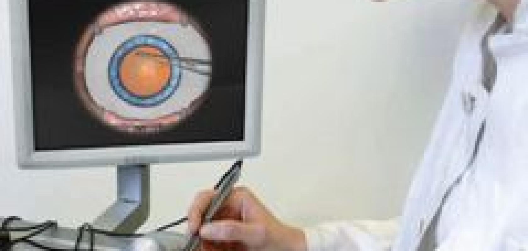
VRmagic, based in Mannheim, Germany, provides training simulators for ophthalmology, based on its expertise in developing software and hardware for virtual reality applications, using vision technology. VRmagic’s Eyesi eye operation simulator enables prospective eye surgeons to train for operations with no risk to patients. It simulates all human pathologies – including rare conditions – and the complexity of the procedure can be adjusted according to the individual’s abilities.
When using the simulator, the surgeon sits in the normal position at an operation microscope and guides freely movable operation instruments into the mechanical eye of a model head. Inside the model head is an optical tracking system that follows the movements of the instruments and transmits their position, alignment and orientation to a computer. The behaviour of fluids and tissue when touched by the instruments is simulated in real time. The operator sees the simulation in the microscope’s displays instead of a real image, with a time delay in visual reproduction of less than the human perception threshold of 50 to 100 milliseconds.

JAI’s AT-200GE Medical, which has colour management qualities making it particularly suitable for use in the medical market
Optical tracking inside the patient’s model head is carried out by means of colour markers: both the pivoted mechanical model eye and the tips of the operation instruments are marked by colours. This means that both the position and the alignment of the eye and instruments can be determined. For the camera system, VRmagic uses a multi-sensor FPGA camera with four offset global shutter sensors, which supply pixel-synchronous images. Following parallel pre-processing of the image data on the camera’s FPGA, the loss-free compressed data stream can be transmitted via a single USB cable to the computer where the data stream is evaluated further. Blob segmentation provides the 2D coordinates for every colour marker, so that the precise location and alignment of the model eye and instruments can be reconstructed in three dimensions.
Based on the measured 3D data, the biomechanical simulation algorithms developed for Eyesi can calculate in real time the way in which tissue behaves when it collides with instruments. The simulated image of the eye’s interior is displayed for both the left and right eye on an OLED microdisplay in the simulator’s stereo microscope. The operator therefore obtains the usual stereoscopic image impression. The update rate of the system is more than 30Hz. The uninterrupted display produces the so-called immersion effect, in which the user no longer perceives the difference between reality and simulated images.
Dry eye syndrome
Vision technology is being used to achieve accurate diagnosis of dry eye syndrome (or keratoconjunctivitis sicca), the fifth largest eye problem in the world, and one that is most prevalent in Asia. The condition means the eyes do not make enough tears, or the tears evaporate too quickly. As a consequence, the eyes dry out or become inflamed. It tends to affect people above the age of 60, and although not usually a serious condition, it can be uncomfortable and, in some cases, can affect vision.
Around four years ago, two Japanese doctors (Dr Reiko Arita and Dr Shiro Amano) began a collaboration with TopCon to develop a vision-based process that would help diagnose the cause of dry eyes, and therefore assist in deciding the most appropriate course of treatment.
Often, the root cause of dry eyes can be traced to the meibomian glands, which are located on the inside of the eyelids; there may be more than 60 of these glands in each eye. The meibomian glands produce lipids, which are an essential component in keeping the surface of the eye moist. If the lipids produced are too ‘sticky’, they cannot get out of the gland. Also, the opening of the gland may be too small.


Using TopCon’s system, medical teams can spot differences between normal meibomian glands in the eyelid (top) and abnormal ones (bottom)
Henry Claessens, product manager at TopCon, explains: ‘In 2009, TopCon Tokyo developed a system using a Sony infrared camera and infrared illumination, which enabled medical teams to see the condition of these meibomian glands. We’ve spent the past couple of years modifying the system for use in the European market. The system uses a slit lamp, which is a biomicroscope specially adapted for viewing the eye by means of a very narrow illuminated slit. It works like an optical knife, slicing through the cornea and into the eye.
‘We’ve been using Sony’s analogue infrared cameras for a long time in our retina imaging systems, so we were familiar with the camera’s quality and also the ease with which it connected to our software.’
TopCon’s system first makes the meibomian glands visible, allowing medical teams to view the exact nature of the condition. ‘We are looking to see if the glands are blocked,’ says Claessens, ‘or if the mouths of the glands are too small. If neither of those appears to be the problem, we look for the presence of the tear meniscus, which will tell us whether or not there is a problem with the aqueous layer on the eye. Once we’ve checked all those conditions and seen no problems, we can deduce that the cause of the dry eyes lays in the absence of necessary proteins in the eyes. At the moment, we can’t yet measure these proteins in isolation, although research is going on to make that possible.’
From this, treatment is then offered, which can include heat treatment if the issue is sticky lipids, or the use of a device to open the mouths of the glands further. ‘The system is used again a few months later to check on the progress of the treatment,’ says Claessens.
TopCon’s system was recently unveiled to the market, and Claessens has been delighted with the response. ‘We’ve been overwhelmed with enquiries, so we are struggling to find the necessary parts to fulfil the orders,’ he says. ‘And following feedback from that launch, we are looking to improve the image quality that the system provides, as this will really help with diagnosis.’
Blood analysis
The use of colour has also had an impact on medical imaging applications, particularly in microscopic applications, such as blood analysis. JAI has recently introduced the AT-200GE Medical, a new 3-CCD digital progressive scan camera aimed specifically at this market. It has been developed to provide accurate, high resolution images under low light conditions, and is particularly relevant to blood analysis, because of its colour management capabilities.
It features approximately five times the resolution compared with ordinary 3-CCD analogue PAL cameras often used in the medical field, and the precisely-aligned three CCDs on the prism block generate true image colours through JAI’s original colour matrix circuit. Users have the possibility to obtain not only the RGB digital data, but also sRGB or Adobe RGB video directly from the camera to calibrate it with connected devices like monitors or printers.
‘The camera features true RGB,’ says JAI’s Michael Lund. ‘Standard colour cameras use interpolation, whereas this unit features true colours, which can be very important in medical applications. The portability of these true colours is also important, as it ensures no loss of colour quality between devices.’
JAI’s true colour management system in combination with Megapixel resolution gives clear and natural images of skin, blood, eye iris and other biological objects being viewed, providing users with a better foundation in relation to patient diagnostics and patient treatment, as well as in medical research studies.
At the Vision show in Stuttgart, several companies will be participating in a new feature called the Medical Discovery Tour, aimed at visitors from the medical device industry interested in machine vision applications. A special guided tour will lead visitors through the exhibition highlighting products and applications, while several presentations during the Industrial Vision Days talks will also link into the medical theme.
