At CLEO 2014, taking place 8-13 June in San Jose, California, scientists from the University of Texas Health Science Center (UTHealth) at Houston Medical School will be presenting a new technology, known as near-infrared fluorescence lymphatic imaging (NIRFLI), which is capable of non-invasively imaging the human lymphatic system using a dye as well as laser and military-grade night vision technologies.
The device is capable of imaging even tiny vessels within the lymphatic system, which has not been possible until now, and will result in earlier diagnoses and improved outcomes for patients suffering from lymphatic diseases.
The human lymphatic system is a poorly understood circulatory system consisting of tiny vessels spread throughout the body. This ‘drainage’ network helps guard against infections and prevents swelling, which can happen when disease interrupts its normal function. For example, chronic swelling is the hallmark of a painful, incurable condition known as lymphedema, which often occurs after cancer therapy and can leave the limbs and other body parts disfigured for life.
Detecting lymphedema early, before swelling occurs, would lead to better outcomes for patients, but the major barrier preventing early diagnosis is the lack of high-resolution imaging techniques that can resolve these tiny vessels. The new imaging technology developed by UTHealth researchers consists of a fluorescent dye and commercially-available laser diode and military-grade night vision devices to visualise the lymphatic capillaries.
Clinically, the device promises dramatic improvements in patient care because it allows even tiny lymph vessels to be imaged, and it can quantitatively measure fluid flow throughout the lymphatic system − two types of measurements that are impossible with today's technology.
At CLEO 2014, UTHealth scientist John Rasmussen will describe how they have taken this technology from bench-top development to various clinical applications. ‘We feel that the ability to see the lymphatics will provide opportunities to revolutionise lymphatic care,’ Rasmussen said.
The major problem with lymphatic imaging is that the small lymphatic vessels are filled with lymph, a clear liquid that lacks the natural contrast needed to show up on instruments like CT scanners or MRIs. And, the lymphatic vessels are very difficult to find and are most often too small to insert a needle, therefore making it almost impossible to inject dyes or other contrast agents.
A technology called lymphoscintigraphy can take images of the lymphatic system following injection of a radioactive compound into or below the skin. However, lymphoscintigraphy typically takes 20-45 minutes to acquire a single grainy picture, and can only image the largest lymphatic vessels or trunks. The smaller vessels, which make up the bulk of the lymphatic system, are invisible to lymphoscintigraphy. In addition, because of the long acquisition times, it cannot capture the real-time flow of fluid in the system.
At CLEO 2014, Rasmussen will describe how near-infrared fluorescence laser imaging (NIRFLI) allows lymphatic structures and flow to be measured quantitatively, including in the fine vessels − a dramatic improvement over lymphoscintigraphy. In addition, NIRFLI will be safer and less expensive than existing technology.
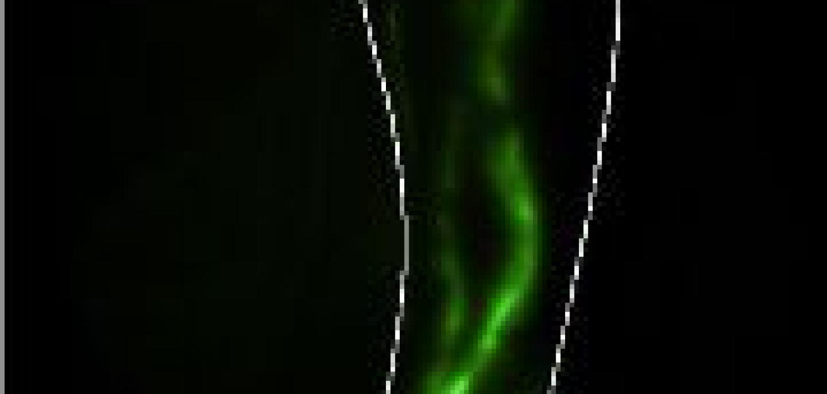
To acquire images of the lymphatics, NIRFLI uses indocyanine green dye, which is injected in tiny amounts into the skin of a patient. The dye is absorbed into the lymphatics and when illuminated by the laser diode, it emits a fluorescent light, which the device amplifies with an image intensifier − the main component in night vision goggles − and then captures with a commercial CCD digital camera.
The image intensifier enables the small lymphatic vessels to be visualised, and by taking sequences of such images, they can produce movies showing flow within the lymphatics. Rasmussen stated that the most immediate promise of NIRFLI will be to diagnose and monitor the treatment of lymphedema and may also help surgeons identify and remove lymph nodes into which cancer tumours drain.
‘From these images and movies, we can identify abnormal lymphatic structure and function in a variety of diseases and disorders in which the lymphatics play a role,’ Rasmussen said. ‘I think we have barely scratched the surface of what is possible.’
The presentation, titled ‘Clinical Translation and Discovery with Near-infrared Fluorescence Lymphatic Imaging', will take place 9 June, at 8am in the Willow Glen I – III Room of the San Jose Marriott.
