Artificial intelligence (AI) is being combined with infrared imaging to optimally tailor colon cancer therapy to individual patients.
The new approach, under development by researchers at the Centre for Protein Diagnostics (PRODI) at Ruhr University Bochum, Germany, is a label-free and automatable method that can complement existing pathological analyses. It has been reported in the European Journal of Cancer.
Immense progress in the field of therapy options over the past years has significantly improved the chances of cure for patients with colon cancer. However, these new approaches, such as immunotherapies, require precise diagnosis so that they can be specifically tailored to the individual.
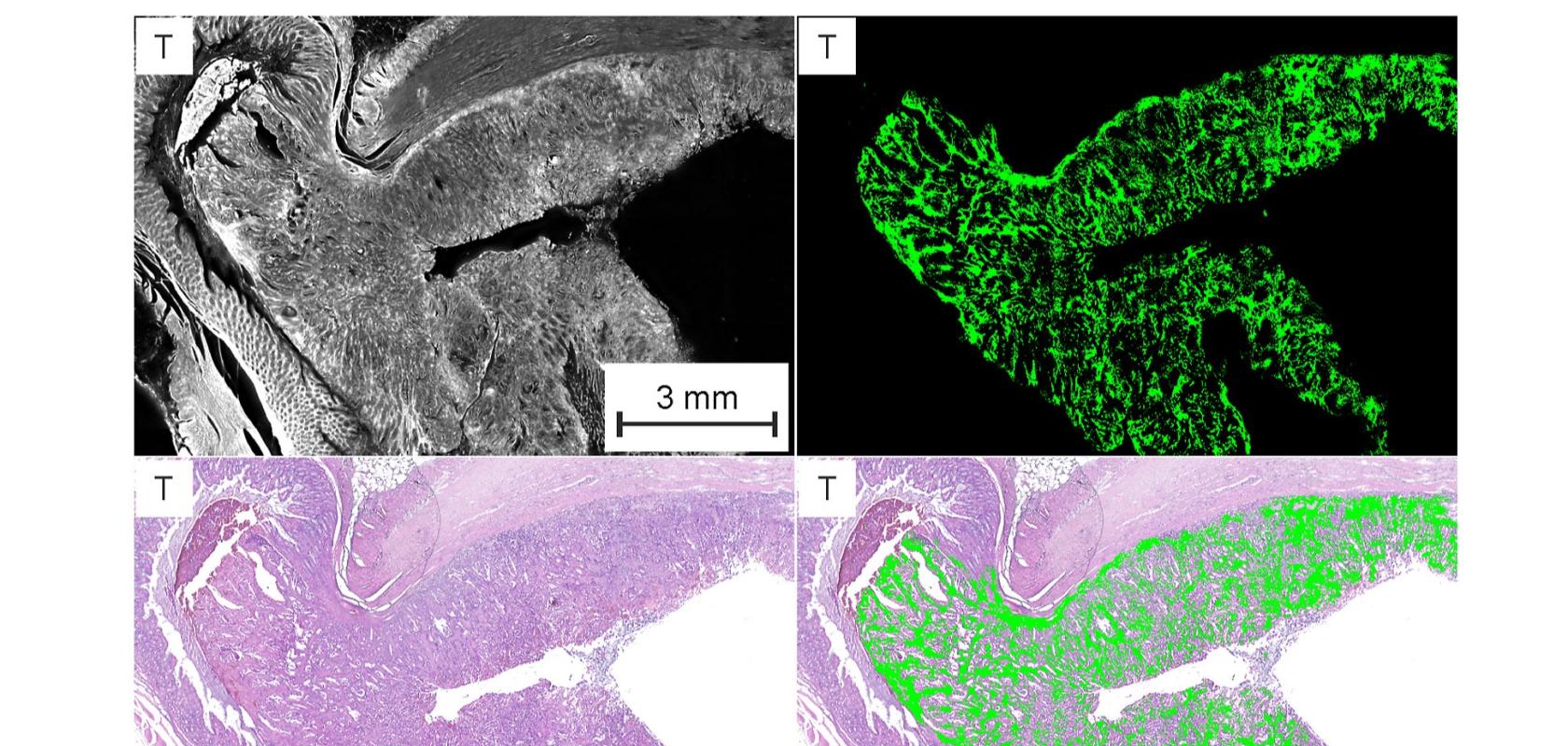
The PRODI team has therefore developed a new AI-assisted, label-free infrared (IR) imaging method to measure the genomic and proteomic composition of the examined tissue, providing molecular information based on infrared spectra. The AI decodes the information – using image analysis methods from the field of deep learning – and displays it as false-colour images.
In cooperation with clinical partners, the PRODI team was able to show that the use of deep neural networks makes it possible to reliably determine the so-called microsatellite status, a prognostically and therapeutically relevant parameter, in colon cancer.
“15-20% of colon cancer patients show microsatellite instability in the tumour tissue,” said Professor Andrea Tannapfel, head of the Institute of Pathology at Ruhr University. “This instability is a positive biomarker indicating that immunotherapy will be effective.”
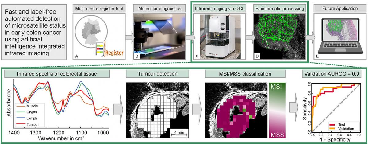
A graphical abstract of the new imaging method. (Image: Gerwert et al./European Journal of Cancer)
In classical diagnostics, microsatellite status is determined either by complex immunostaining of various proteins, or by time-consuming DNA analysis. However, using the new AI-assisted infrared imaging method the tissue sample instead goes through a standardised, user-independent, automated process that enables a spatially resolved differential classification of the tumour within one hour. This is done without the use of dyes and with an accuracy approaching that of immunostaining – currently the most common clinical method. The researchers believe the accuracy of their technique can be increased even further through continual development and optimisation.