CEA-Leti scientists have developed a lensless, infrared spectral imaging system for medical diagnostics. The French research agency has a start-up in incubation based on the early-stage technology.
The technology has been demonstrated in cancer detection in tissue samples, and also in identifying and discriminating between species of bacteria. The group says the label-free technology could eliminate sample preparation and therefore lead to greater automation in diagnostics.
The work was presented during SPIE Photonics West, which took place last week in a digital format.
The scientists showed that the mid-wave infrared multispectral imaging method could detect cancer more accurately and faster than a human assessing a tissue sample, which is the standard practice for identifying cancer.
The paper reported that by analysing images from mice tissue using amide and DNA absorption bands, the team 'achieved up to 94 per cent of successful predictions of cancer cells with a population of 325 pixels corresponding to muscle tissues, and 325 pixels corresponding to cancer tissues. This work may lead to the development of an imaging device that could be used for cancer diagnosis at hospitals.'
'Employing recent developments in photonics components, which allow using infrared light to detect abnormal tissues, mid-infrared imaging can provide unequivocal information about the biochemical composition of human cells,' said Grégoire Mathieu, lead author of the tumour detection paper. 'The combination of a set of lasers and lensless imaging with an uncooled bolometer matrix allows biochemical mapping over a wide field of view. The project showed that this experiment’s setup coupled to machine learning algorithms – Random Forest, Neural Networks, K-means – can help to classify the biological cells in a fast and reproducible way.'
The second technique is an optical-based, Petri-dish analysis using lensless multispectral mid-infrared imaging. Described in a paper, 'the technique relies on the acquisition of images at eight wavelengths corresponding to relevant chemical functions. It provides both morphological and discrete spectral data, which discriminates between even closely related species.'

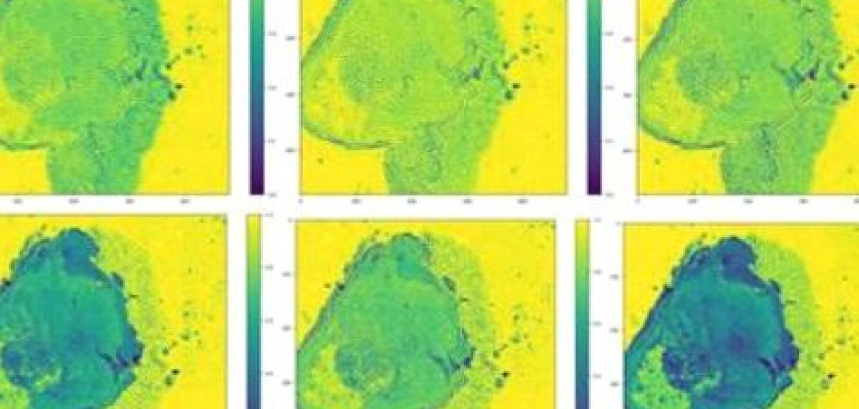
Multispectral images of representative examples from the seven species of the database. Wavenumbers on top of each column are in cm-1. Credit: CEA-Leti
For this proof of concept, a database containing 2,253 colonies belonging to eight different species and three strains of Staphylococcus epidermidis was acquired. The optical setup and machine-learning analysis could classify all species with a correct identification rate of at least 91 per cent.
The early-stage technology used in both studies was enabled in part by recent improvements in photonics components at CEA-Leti. The next steps are to perform a dedicated prototype with the relevant wavelengths and to demonstrate the performance of the system with real-life samples, such as human biopsies, and to create larger databases for each application.