Non-invasive breakthrough in ear diagnostics uses terahertz imaging
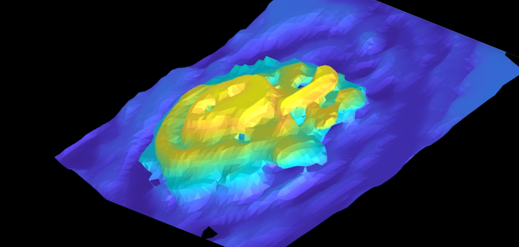
Images acquired using 3D terahertz near-field imaging were used to create 3D reconstructions, allowing visualisation of part of the cochlear duct, the spiral structure inside the cochlea. (Image: Kazunori Serita, Waseda University)
Terahertz imaging has been used to offer the precise and non-invasive visualisation of the cochlear structures of mice in a study on ear diagnostics
Register for FREE to keep reading
Join 10,000+ vision professionals driving innovation in automation, AI and imaging with:
- Expert insights on vision, robotics, AI & embedded tech
- Newsletters and features covering the full imaging landscape
- Visionaries series: leadership strategies in imaging
- Free panels on smart manufacturing & autonomy
- White Papers & updates for smarter integration
Sign up now
Already a member? Log in here
Your data is protected under our privacy policy.