Vision Standards Bring Sharper View to Medical Imaging
The noisy factory floor may seem worlds away from the sterile hum of a hospital operating room, but the inspection cameras and robotic arms along a manufacturing line are played an important role in the continuing evolution of medical imaging. This paper discusses how vision standard-compliant GigE and USB 3.0 video interfaces widely adopted in industrial automation applications are supporting the development of more sophisticated, easier-to-use medical imaging systems.
GigE Vision and the Networked Operating Room
Medical imaging systems require video interfaces that reliably transfer high-resolution images from cameras or image sensors to computers and displays in real-time with low, consistent latency (or delay). Fluoroscopy, for example, requires fast transfer of images from a flat panel detector (FPD) to a display screen to enable precise surgical decisions. Beyond technical performance, new systems must integrate seamlessly with existing equipment and scale easily to add additional image sources or display panels.
The GigE Vision standard is a good starting point to meet these requirements. With this standard, full resolution uncompressed video is transmitted with low, consistent latency directly to an existing Ethernet port on computers used for analysis, display, and recording. This eliminates the need for costly PCIe frame grabbers to capture images at endpoints, while enabling the use of a wider range of computing platforms including laptops and tablets.
The same Ethernet connection transmits control data between the computing platform and the imaging device, as well as configuration information for imaging systems used for different procedures. Per-frame metadata, such as the date and time of acquisition, sensor settings, and imaging equipment used, is also transmitted over the GigE link for easy integration with DICOM-compliant software and hardware.
Thanks to the widespread adoption of the GigE Vision standard in the industrial sector, compliant products are widely available as off-the-shelf solutions that make it relatively straightforward for designers to create a real-time networked medical imaging system.
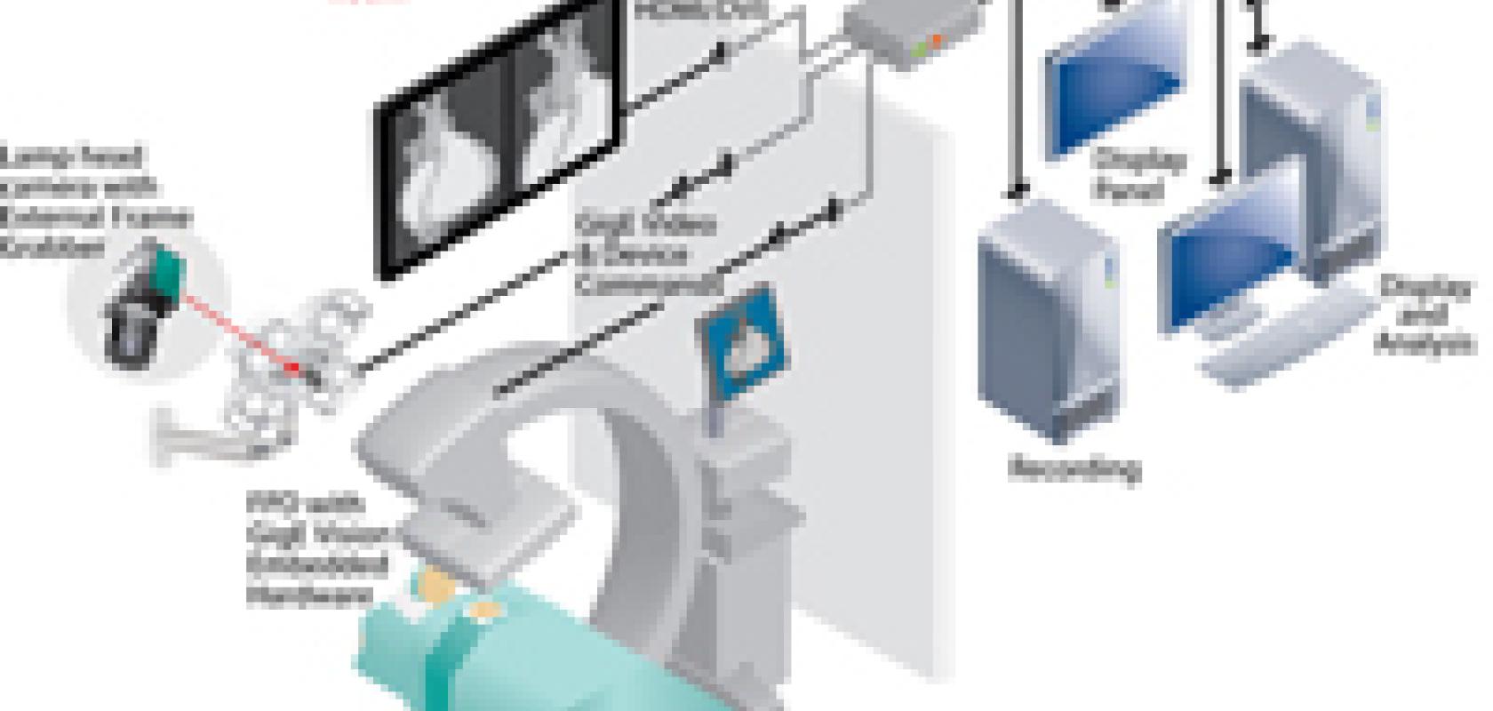
In a networked operating room (Diagram 1), a film-based X-ray panel in the C-arm has been replaced with a digital FPD. Embedded video interface hardware allows designers to easily integrate GigE Vision-compliant video connectivity into FPDs, with numerous manufacturers now offering digital panels as a direct drop-in replacement for film-based panels. In systems where images are being sent from a FPD over an existing legacy interface, such as Camera Link or LVDS, an external frame grabber can convert images into a GigE Vision-compliant video stream. Image feeds from a lamp head camera used to observation and archiving are also converted into the GigE Vision format by an external frame grabber.
Diagram 1: GigE Vision video interfaces for medical imaging system help preserve capital investments in cameras, sensors, and processing systems — while enhancing outcomes — by enabling networking and cabling advantages.
Image feeds from the FPD and camera are aggregated at an off-the-shelf Ethernet switch and multicast to video processing, analysis, and display equipment. The long reach of Ethernet – 100 meters over Cat 5/6 cabling – means processing and analysis equipment can be located outside the sterile environment.
The video processor creates a composite image, highlighting areas of interest and overlaying pre-op images and vital signs information, that is then multicast over the Ethernet network to various displays. In the operating room, an external frame grabber converts the GigE Vision image stream to HDMI/DVI signals for viewing on a high-definition dashboard.
One of the key performance advantages of the GigE-based distributed network architecture is the ability to aggregate and display information on a single dashboard. In the operating room, for example, the single screen dashboard displays recorded and real-time patient data from different imaging devices and systems. The surgeon or operating team members can easily switch between imaging sources, such as white light and fluoroscopic cameras or pre-op and real-time images, without reconfiguring hardware.
Increasingly, designers are seeking wireless solutions to enable more portable imaging systems. GigE Vision over an industry-standard 802.11 wireless link sets the stage for untethered imaging systems that can be better positioned for patients with limited mobility. Portable imaging equipment can also be conveniently located in a crowded OR to help improve room management.
Plug-and-Play Advantages of USB3
While USB is one of the most popular interconnect approaches for consumer electronics, its use in medical applications had primarily been limited to home-based health monitoring devices. This changed with the release of the “super speed” USB 3.0 and development of the USB3 Vision standard.
With USB3 Vision, data is transmitted directly to existing ports on a computer with sustained throughputs approaching 3 Gb/s. This surpasses the performance of Camera Link Base configurations and rivals Medium configuration, but without requiring specialized frame grabbers at endpoints to capture data to enable the use of a wider range of computing platforms. Beyond speed advantages, the simplified cabling and plug-and-play usability of USB 3.0 delivers cost, performance, and reliability advantages in a wide range of medical imaging applications.
In microscopy applications (Diagram 2), an external frame grabber allows designers to retain existing cameras, sensors, and optics while leveraging the ubiquitous computing platform support and simplified cabling of USB 3.0.
Microscopy systems often use a Camera Link camera to transmit microscopic images to a computer for analysis and observation. With an external frame grabber, the image feed from a Camera Link microscopy camera can be converted into USB3 Vision-compliant video. The uncompressed video is transmitted with low, consistent latency over a USB 3.0 cable directly to an existing USB 3.0 port on a laptop used for analysis and display. The thinner, lighter USB 3.0 cable is easier to route than bulky Camera Link cables and connects with “plug-and-play” ease, allowing faster setup and teardown of inspection stations.
Diagram 2: USB3 Vision external frame grabbers reduce cabling complexity and system costs in microscopy applications by treating Camera Link cameras like plug-and-play USB 3.0 products.
Increasingly, robotics manufacturers are migrating to embedded processing to help deliver footprint, weight, and power-saving advantages. However, as form factors shrink there is no room for an internal frame grabber. USB3 Vision external frame grabbers or embedded hardware can be deployed to simplify the design of robots used for medical service or telepresence applications (Diagram 3).
In this example, images from cameras used for inspection and navigation in a medical service robot are converted to USB3 Vision-compliant video streams by external frame grabbers. Alternatively, embedded video interface hardware provides system and camera manufacturers with a straightforward way to integrate USB 3.0 video connectivity directly into their products.
Video, power, and control data is transmitted over high-bandwidth, flexible, low-cost USB cables directly to ports on an integrated single-board computing platform. By eliminating PCIe frame grabbers within the robot, designers can reduce system complexity, component count, and costs. In addition, decreasing the weight and power consumption of the robot extends battery life, translating into more patient visits between charges.
Diagram 3: USB3 Vision-compliant external frame grabbers or embedded hardware simplify the design of robots used for medical service or telepresence applications.
New Levels of Performance
Medical imaging technology is enabling new levels of precision, insight, and diagnoses to improve patient outcomes and reduce costs. Many of the benefits offered by these imaging systems can be traced back to the choice of video interface. Designing or upgrading medical imaging systems with off-the-shelf vision standard-compliant video interfaces allows manufacturers to shorten time-to-market, reduce risk, and lower system cost and complexity, while delivering interoperability and performance benefits to enhance the value of their solutions.
