Researchers from North Carolina State University and the University of North Carolina at Chapel Hill (UNC) have developed a new tool to help surgeons use X-rays to track devices used in 'minimally invasive' surgical procedures – while also limiting the patient’s exposure to radiation from the X-rays.
Many surgical procedures now use long, thin devices – such as 'steerable needles' – that can be inserted into a patient’s body through a small incision and then steered to a target location. These 'minimally invasive' procedures allow doctors to perform surgeries without having to make major incisions, which decreases the risk of infection and shortens recovery time.
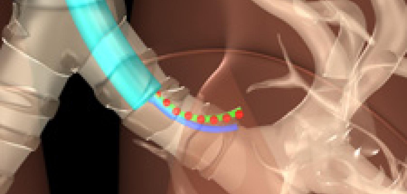
However, these techniques pose a challenge to surgeons, because it is difficult for them to determine precisely where the surgical device is in the patient’s body. One solution is to use X-rays to track the progress of the surgical device in the patient – but doctors want to minimise the number of X-rays taken.
'We have now developed an algorithm to determine the fewest number of X-rays that need to be taken, as well as what angles they need to be taken from, in order to give surgeons the information they need on a surgical device’s location in the body,' said Edgar Lobaton, assistant professor of electrical and computer engineering at NC State and lead author of a paper on the research.
The new tool is a computer program that allows surgeons to enter what type of procedure they’ll be performing and how precise they need the location data to be. Those variables are then plugged into the algorithm developed by the research team, which tells the surgeon how many X-rays will be needed – and from which angles – to produce the necessary location details.
For example, if a surgeon needs only a fairly general idea of where a device is located, only two or three X-rays may be needed – whereas more X-rays would be required if the surgeon needs extremely precise location data.
