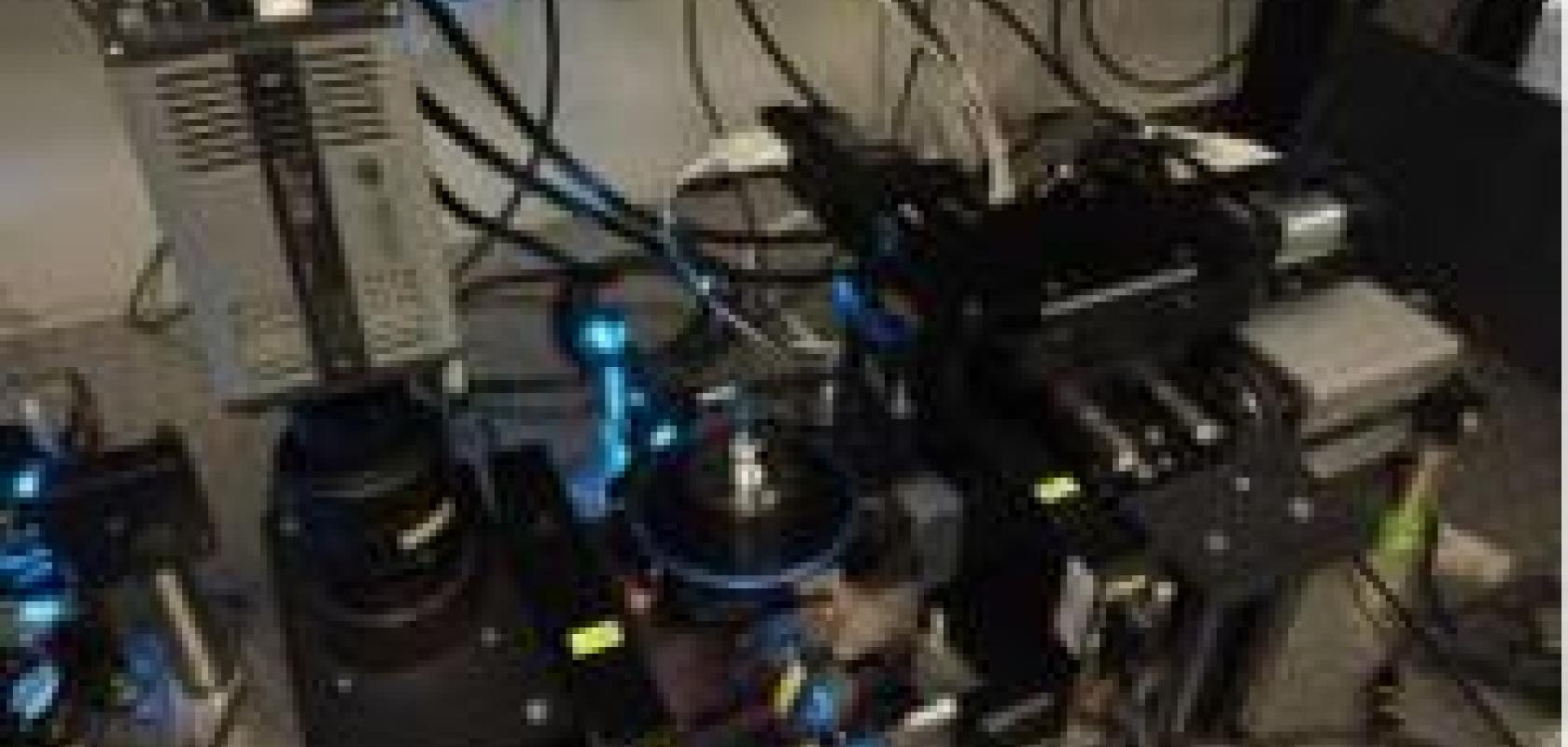
A team of researchers from the Max Planck Institute of Molecular Cell Biology and Genetics and the Technical University in Dresden, Germany, has created a specially designed four-lens selective plane illumination microscope (SPIM). Containing two Andor Zyla sCMOS high-speed cameras and on-board image processing, the microscope can deliver undistorted, high-resolution images of the entire endoderms of multiple zebrafish embryos in less than ten seconds.
Light-sheet microscopy is rapidly gaining recognition for whole-organism imaging, allowing cell growth, differentiation and morphogenesis to be studied in detail. However, the technique generates approximately three thousand times as much data as confocal microscopy and this amount of data poses challenges in storage and the post-processing of images.
Rather than deploying greater storage and computing power, the team from the Max Planck Institute and the Technical University has reduced the data rate 240-fold to create the new SPIM that processes image data in real-time and provides the researcher with results rather than raw data.
Jan Huisken, research group leader at the Max Planck Institute, explained: ‘An entire zebrafish embryo can now be instantaneously projected onto a "world map", to visualise all endodermal cells. By merging data from many samples, we have uncovered stereotypical patterns that are fundamental to embryo development.
‘The raw data from the Zyla cameras was not saved at any point, meaning that 3D information not captured in the projection is lost,' Huisken continued. 'However, the radial projection delivers immediate, pre-processed data for analysis, and enables experiments to be repeated rapidly. This technique is an effective strategy to streamline further analysis and increase throughput in many applications.’
Orla Hanrahan, application specialist at Andor, said: ‘Image processing in real-time is a major advance for researchers who recognise that statistical, large-scale analysis is impossible when single experiments produce approximately 13Tb of data. Jan Huisken's microscope reduces the data collected so that many experiments can be performed. The data is projected in a convenient way to facilitate further analysis, and this principle can also be used to address other questions in developmental biology.’


