Since its emergence in Mexico, swine flu has spread across the globe, with the World Health Organisation (WHO) officially declaring it a pandemic on 11 June 2009. As of 31 July 2009, 168 countries and overseas territories have reported at least one laboratory confirmed case of the pandemic H1N1 2009 strain and a total of 162,380 cases have been reported, according to WHO figures.
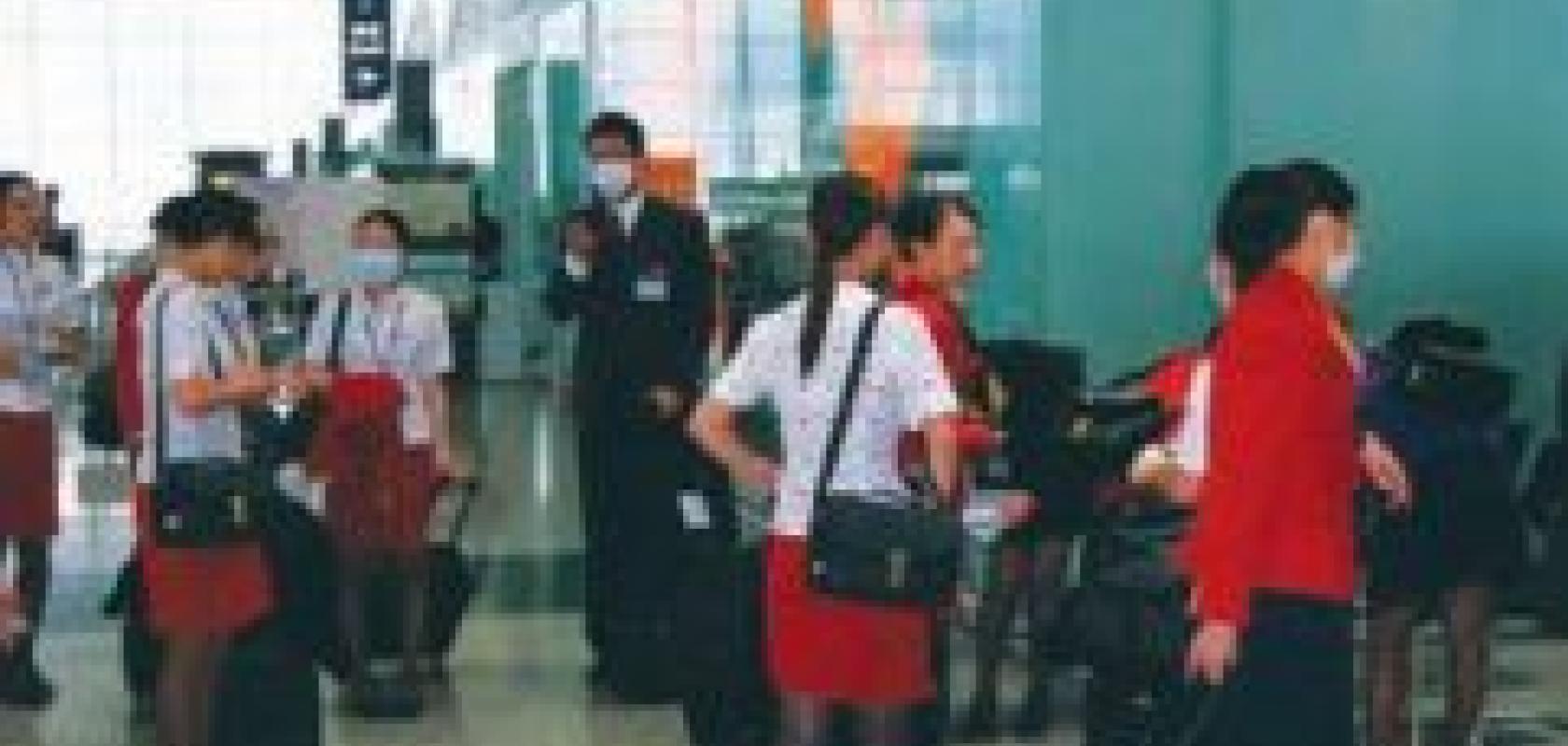
The symptoms of swine flu are similar to other seasonal flu strains, the main one being a fever (a temperature of 38°C or higher), with other complaints, such as a cough, sore throat, body aches and chills, common among sufferers. Thermal imaging was first used to screen people for signs of fever on a large scale during the 2002-3 SARS (severe acute respiratory syndrome) outbreak; airports in Asia screened travellers in an effort to contain the spread of the disease. With the recent swine flu outbreak, there is increased interest in utilising thermal imaging cameras for fever screening.
The technology
Thermal imaging cameras detect variations in infrared radiation, based on an object’s temperature (according to Planck’s law), all objects radiate energy according to a given theoretical curve. Thermal imaging sensors are typically focal plane arrays (FPAs), which measure a change in resistance on each pixel when exposed to infrared radiation – as opposed to photon detectors, which detect photons of light. Thermal cameras operate over the mid (3-5μm) to long (7-14μm) wave infrared wavelengths.
Thermal imaging cameras are split between cooled and uncooled technology. ‘Ten years ago, the majority of thermal imaging cameras were cooled, in that the sensor was cryogenically cooled to around -190°C,’ remarks Paul Sacker, sales and marketing manager at Flir Systems, a manufacturer of thermal imaging infrared cameras. Older models used liquid nitrogen as a coolant, while today’s models typically use a stirling engine cooler.
Cooled cameras provide good signal to noise ratio and therefore high sensitivity, detecting temperature differences down to 0.015°C. However, the cameras are expensive and, as they are mechanically cooled, have a limited life. Typical applications for cooled cameras include high-end, military and defence R&D, such as target signature analysis or detecting warm objects over large distances. Uncooled camera technology, on the other hand, is easier to make, cheaper, and the run time is not limited. The sensor arrays are commonly fabricated from amorphous silicon or vanadium oxide. The sensitivity is less than cooled cameras – the smallest detectable temperature difference is around 0.05°C – but this is more than adequate for many applications.
Jean-Luc Tissot, technical director at infrared detector manufacturer ULIS, makes the point that advancements in detector fabrication techniques are allowing the production of smaller pixel sizes. ‘Smaller pixels allow for an increase in resolution and a reduction in the size of the system,’ states Tissot. The size of pixels in ULIS’ thermal imaging detectors has decreased from 45μm to 35μm in 2003 and then 25μm in 2005. This is to decrease further to 17μm and the company is to release a 640 x 480 format of its 17μm pixels IR detector later in the year.
Fever screening
‘The relative cheapness of uncooled cameras, compared to cooled technology, makes thermal imaging accessible for an increasing number of applications,’ comments Sacker. In the case of fever screening, cameras can be set up to monitor people as they pass through airports or other major transport hubs. In principle, the system will alert officials, either via a visible or audible alarm, if a person’s temperature exceeds a set limit – typically 38°C – an indicator that the person is suffering from fever.
The most reliable indicator of body core temperature on the face’s surface is the inner canthus of the eye (the inner corner of the eye) and this is where temperature measurements should be made if the fever screening system is to be effective. The inner canthus is fed by the internal carotid artery with the circulation coming directly from the brain, whereas the rest of the face is supplied by the external carotid artery and is therefore a less reliable indicator of body core temperature.

Thermograms of the face showing a healthy individual (left) and a fever sufferer (right). Image courtesy of the Medical Imaging Research Unit (Prof FJ Ring), University of Glamorgan.
Professor Francis Ring of the Medical Imaging Research Group, University of Glamorgan, and part of the ISO standard writing group on thermal imaging for fever screening, states: ‘To obtain an accurate measurement, the person’s face should be looking straight into the lens and the camera should be close enough to obtain a minimum of 16 pixels in each corner of the eye.’ Spectacles, facemasks, baseball caps pulled over the eyes – all must be removed to get an accurate reading. The exact image capture procedure, which was developed at the University of Glamorgan, has been adopted into the international standard for fever screening.
According to Ring, one of the disappointments from the SARS material is that, although a lot of anecdotal information came out of it, very few documents were kept or published. The criterion used was simply to put a threshold of 38°C on the images and have an alarm system when the temperature of the face exceeded that level.
‘The technique was based on the premise that somebody with SARS had a very hot face, so that if they were walking in a crowd, a thermal camera could pick them out. The problem is, that this is not really the true situation with the influenza virus,’ Ring says. ‘In extreme cases of fever the whole face will be hot, but if a person’s that sick they probably won’t be travelling.’
Two standards have been established detailing design and performance specifications of the cameras and how they should be employed, tested, and maintained, as well as any staff training requirements. ‘One of the things the standard requires is that the equipment is treated as a medical device and that means certain specifications have to be adhered to,’ comments Ring.
Criteria that the standard recommends for effective fever screening include: the camera should reach stability prior to use; equipment should be calibrated frequently; the alignment of the camera with the face is important; and cameras should be checked regularly and the data backed up.
One of the criteria the standard recommends is that, with an uncooled camera, a temperature reference source is included in the scene, so each image is in effect carrying its own calibration. ‘Making a decision on whether a person is running a fever or not requires an accurate method for temperature measurement and including a reference in the frame maximises the accuracy of the reading,’ remarks Martin Ghillemyn, sales Europe at Xenics. Xenics’ Raven-384 uncooled microbolometer camera is suited to fever screening.
Calibration of infrared cameras is typically carried out under rigidly controlled environmental conditions. At Xenics, this is done in a climate chamber using high-standard black body references. This is especially important with uncooled bolometer cameras, as the sensor’s sensitivity is affected by environmental conditions. Despite in-house calibration, a black body reference, placed in the background of a passenger checkpoint, is still recommended to obtain an accurate reading.
Accuracy is key
There are still unknowns about the H1N1 virus itself, such as the time course of reaching fever point. This can vary, with some people reaching fever point very quickly – in a matter of hours – while, for others, it could take days. ‘There are always going to be imponderables; we don’t know everything there is to know about the course of fever, but the best we can do is to apply the technology as accurately as possible,’ says Ring.
Sacker, of Flir, comments that the system can produce false positives: ‘An elevated body temperature can be caused by variables other than viral infection – for instance, people running for their flight or people drinking hot beverages will be warmer than otherwise.’ However, it’s better to have a screening system – if the proper equipment is used and the system is set up correctly – than none at all.
Sacker also notes that businesses have also shown interest in installing thermal imaging equipment to minimise the spread of the H1N1 virus within office buildings and maintain a healthy workforce.
Over the last four years, Ring has been carrying out studies at the Military Institute of Medicine in Warsaw, where febrile children have been screened using thermal imaging. ‘The conclusion drawn is that the technique definitely does work,’ he says. ‘There was a high correlation between body core temperature and measurements from thermal images of the eyes, but that’s when the subject is looking straight into the lens using a high-quality image.’
While thermal imaging cameras are in high demand for fever screening applications due to the current swine flu pandemic, there are a host of other applications where the technology is used. Military and defence is an obvious big user, but the cameras are also used in medical settings, as a diagnostic technique for circulatory disorders; in building inspection, to identify areas of heat loss; by firefighters, to see through smoke; and in the analysis of art, to look through layers of paint.

Automotive manufacturers are developing vehicle models that use thermal imaging as a driving aid. Image courtesy of ULIS.
Flir Systems’ cameras are used in a project to determine the health of trees. ‘Amenity and leisure services can make use of the technology to identify deadwood and determine whether individual branches need to be lopped off or the entire tree cut down,’ explains Paul Sacker, sales and marketing manager at Flir.
Jean-Luc Tissot, technical director at ULIS, a producer of infrared detectors, notes that car models are being developed that use thermal imaging cameras as a driving aid. The technology is designed to increase the driver’s awareness of their surroundings when driving at night or in fog. ‘Because infrared vision is a passive technology, the camera will cut through the fog and show any warm objects in the vehicle’s path, such as a pedestrian,’ explains Tissot. The image will not alter what the driver sees through the windscreen but simply act as an aid in these types of difficult driving conditions.