The use of digital technology in dentistry has been acknowledged for some time as a way to make these procedures more efficient than the more traditional mechanical tools. But, with clinics gradually beginning to invest in this kind of technology – which itself is developing at pace – ‘digital dentistry’ is starting to become more mainstream.
In research and academia the technology is not new. In the early 1970s, Francois Duret was credited with inventing the first computer-assisted design/computer-assisted manufacturing (CAD/CAM) tool for oral healthcare after conceptualising how the kind of 3D digital technology used in other industries could be adapted to this market.
To use a more recent example, Pete Tomlins, senior lecturer in oral biometrics at Queen Mary University of London, is currently undertaking research on using optical coherence tomography (OCT) to diagnose and understand oral diseases of hard and soft tissue, such as oral epithelial dysplasia, periodontal inflammation, caries and erosion. Tomlins is focusing on the translation of his laboratory-based studies into clinical use.
Bit between the teeth
Tomlins explained: ‘Dentistry currently uses lots of x-rays and my interest in is replacing these x-rays with alternative technologies.’ The reason, he continued, is that there is ‘no level of safe x-ray’. The OCT imaging technique can look both inside and between teeth, which is often what a dental x-ray is used for.
X-rays can identify whether tooth decay is beginning in between the teeth, where a mirror cannot reach. If decay is starting, the earliest stage is called de-mineralisation or white spot lesions.
‘The problem with x-rays,’ continued Tomlins, ‘is, for one, they are not really good at seeing any lesions, because all the information is compressed down into 2D.’ In addition, tooth density will vary because of the curvature of the tooth. ‘There are all these confounders that could make a lesion either look smaller or bigger, or it might not even show up at all. Early lesions are quite difficult to spot on a dental x-ray, as they’re not very high-quality images.’
Safe and sound
The other issue is the safety of x-rays, especially when treating children for white spot lesions. Treating the lesions involves monitoring the re-mineralisation process over time, which potentially means exposing the child to more x-rays.
A solution, Tomlins believes, is to use near infrared light to see these lesions. He’s developed a prototype, pre-clinical, laboratory-based imaging instrument, and is applying to Barts Charity for a grant for further work.
When a tooth de-mineralises, some of the enamel crystal structure is broken down and dissolved in the acidic saliva. Enamel crystals are transparent to infrared light, but demineralised areas will scatter infrared.
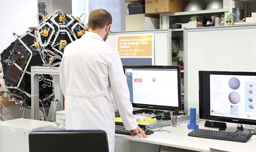
The Zero Gravity 3D system can be used for defect detection. Credit: ITI
‘This allows us to image inside the tooth where these lesions have formed and how deep they have gone,’ explained Tomlins. ‘My work is really trying to create 3D images to measure the depth, or severity, of these lesions in between the teeth, and then provide that as a measure to dentists, rather than using x-rays. And it’s safe. One thing we want to do before going to patients is finalise the protocol for how to measure these things [lesions] in the mouth.’ After this protocol is complete, the technology can proceed to a clinical trial.
One of the challenges of bringing this type of imaging technology to the clinic is trying to drive down the cost for what are fairly expensive pieces of equipment, in comparison with those used for a bitewing dental x-ray. Tomlins will use some of the Barts Charity grant to source lower-cost components, in order to turn a bulky and expensive lab setup into a much smaller bitewing x-ray replacement. He hopes to start moving to clinical work in the next year.
Freeze frame
Moving from diagnosis to manufacturing, the IT Technological Institute (ITI) in Spain has developed the Zero Gravity 3D system, a scanner with a 16-camera configuration to ‘freeze’ parts under inspection. One of the uses for which the system was designed is to measure and inspect dental prostheses.
What makes the system different, according to Professor Juan Carlos Perez, director of the machine learning and artificial intelligence laboratory at ITI, is its use of freefall technology. The objects to be inspected enter the device automatically, by freefall from the top or by being pushed up from the bottom, which allows the object to be captured in its entirety (360°) without the need to manipulate it and without any occlusions.
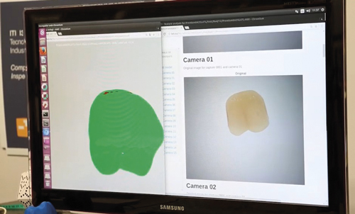
Software digitises and performs the 3D function. Credit: ITI
The 16 cameras are distributed in a spatial arrangement that has been optimised to ensure accuracy. The system is devised to inspect the kinds of components and parts that would typically go on a conveyor and have a robot reach out to grab them. All 16 cameras take an image of the object, which is then reconstructed in 3D in ITI’s software. The surface of the object can then be compared to a reference model for quality assurance.
Towards automation
Focusing specifically on dental prostheses, Sergio Navarro, business development manager at ITI, added: ‘In this case, in particular with dental prosthesis inspection, we work with companies that – based on high level of quality – are currently doing it manually, with several people working in the quality control department. They need, every day, to check each part of their production – both their general quality, but they also need to look for various small defects that can be on the outer and also in the inner layers of each part [dental prosthesis].’
Perez added: ‘Dental layers are fairly transparent; you’ve got to give the impression of a real tooth that has special optical properties to disperse the light and so on, so a defect inside can be miss-able.’ The system can check for colour, scratches and bubbles on the inner layer of prostheses. Breaks and dust can get in the tooth face, in the inner layers, but cannot be removed by cleaning the part, according to Navarro. Manufacturers want to identify defects in the range of 50-100µm, Navarro added; the Zero Gravity 3D system can identify defects as small as 20-50µm.
The system has been in development since 2010. The original need from industry stemmed from the automotive sector, where there was an expectation for manufacturers to develop new systems to control in-line parts such as springs, screws, nodes and other small components. ‘The problem,’ said Navarro, ‘is that these kinds of parts are very cheap, but they need to be able to control 100 per cent of their production – manufacturers request 100 per cent quality control for each part.’
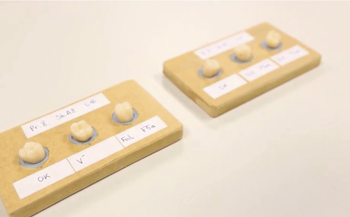
Dental implants can be checked with the Zero Gravity 3D system. Credit: ITI
Perez agreed: ‘They have a high impact on the production and the quality, and if a very simple part breaks or works badly, it can impact on safety.’
Initial feedback indicates that industry likes the system. Navarro said: ‘We have two prototypes in our laboratory, but we are making the first steps in real companies. This year, we started a test in a company near us, from the automotive sector, and at the end of the year we are also looking to start a new test phase with industrial machines with another customer.’ ITI is also working on a speciality dental prosthesis project.
Perfect partner
There has been a lot of interest and the institute is at a stage where it is looking for partners. Navarro said: ‘We have developed the technology and we need to demonstrate in a realistic scenario.’
The team believes the system’s main advantage, aside from the technology, is that it can work with many different parts at the same time, one after the other, without telling the system what’s coming next. ‘It’s very different from what’s now in the industry,’ Perez said. ‘Most of the systems are made for particular parts, so if you make 10 million identical parts a year, you need another solution.’