An unpleasant part of any hospital visit is the multiple needle sticks required when a nurse searches for an elusive vein. Difficulties associated with intravenous access have driven the development of new imaging technologies, such as a pair of 3D glasses, due to enter the market early this year, and a robotic needle-guidance system that will be in the clinic within three years.
Many hospital treatments begin with intravenous (IV) access to allow for sampling of blood, as well as administration of fluids and medication. For IV placement, nurses currently rely on the appearance and feel of veins on the surface of a patient’s skin. ‘In many cases, due to subcutaneous fat, hair, freckles, or darker complexions, nurses can’t see or feel veins – in which case it’s like shooting in the dark,’ said Frank Ball, CEO of Evena Medical. ‘Studies have shown that up to 40 per cent of IV starts require multiple attempts to locate and access a vein.’
Repeated attempts cost time, delay therapy and cause discomfort to the patient. In addition, a misplaced line can cause IV fluid or medications to leak into the arm tissue, which can result in serious complications, particularly for chemotherapy patients. It is because of issues such as these that the Infusion Nurse Society (INS) – the organisation that sets protocol for infusion therapy in the USA – now recommends that IV placements should be carried out with the assistance of either sound or light imaging.
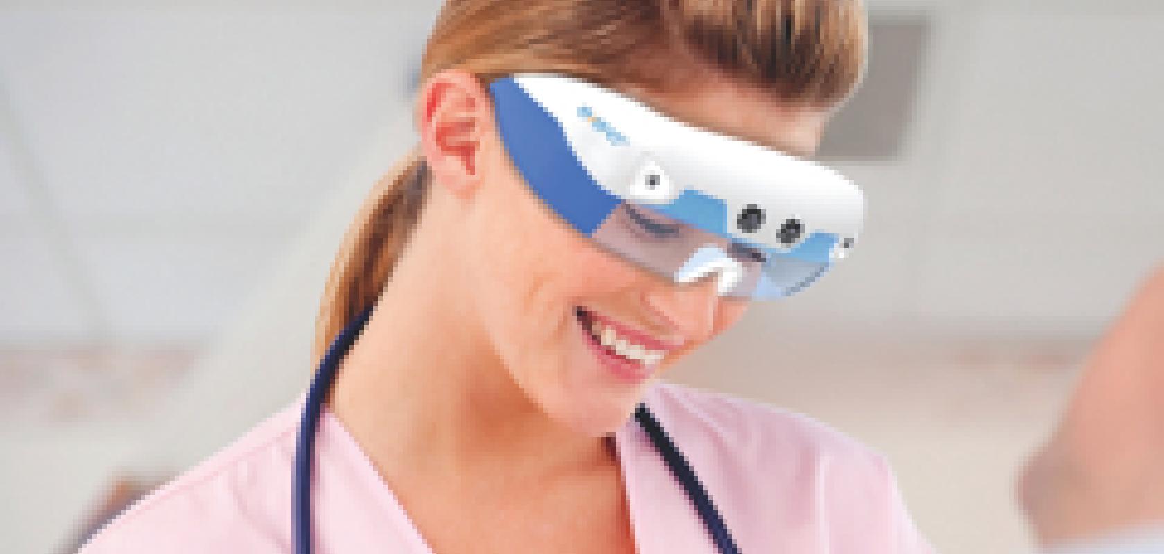
This April, a wireless glasses system will become available which should make IV access for blood sampling or infusing medication an easier process for all involved. The Eyes-On Glasses system developed by Evena Medical in the USA uses 3D imaging to allow nurses to see ‘through’ a patient’s veins, in real time, to locate the best veins for each patient.
Evena’s Eyes-On Glasses system uses near-infrared (NIR) imaging to view the vasculature. Because blood absorbs a high level of light in the NIR range, as compared to other biological tissues such as skin, muscle and water, an optical contrast is created between the veins and the surrounding tissue. By using the system, nurses will also be able to check if IV fluid is leaking out of a patient’s vein over time. ‘We use a higher wavelength than most other vascular imaging systems,’ explained Ball. ‘This allows us to see finer detail, so we can see the blood flow and spot if there is any leakage.’
The system is based on Epson’s Moverio Smart Glasses technology, a see-through wearable display launched earlier this year that allows users to interact with games and apps. Evena’s glasses have multispectral lighting on each side, along with two custom-digital, hypersensitive cameras and two digital light projectors (DLP).
To present the 3D image in a heads-up display, the cameras take two separate images, which are wirelessly transmitted to a processing unit attached to the nurse’s belt, before being sent to the DLPs. The two projectors then fire each image forward independently to create a stereoscopic 3D display directly in front of the practitioner’s eyes. The end result is that the practitioner sees the patient’s skin through the glasses’ clear lens, but with an image of the veins overlaid on top. In addition, the cameras are aligned with the practitioner’s eyes, so that when their eyes move, the projected image will always be in the practitioner’s line-of-sight.
The glasses will arrive amid a flood of wearable computing devices coming to market, including Google Glass, which has received attention in the medical device industry for its ability to film medical procedures. ‘Some people ask us the difference between our glasses and the Google Glass,’ explained Ball. ‘The big difference is that the Google Glass was simply not designed for medical applications.’ For the Eyes-On glasses to be used in a healthcare setting, there were specific factors that Evena had to consider, such as interaction between the healthcare professional and the patient. ‘There are glasses systems out there that have reticules in front of each eye and can create a very nice image,’ explained Ball. ‘Unfortunately, they don’t have any situational awareness – the practitioner wouldn’t be able to have eye contact with the patient.’ Conversely, similar technologies position the image above the eye, to the side of the eye, or in the case of Google Glass, in the upper right hand corner. ‘If this was the case, the practitioner might have the tendency to curve [their movement] to the right,’ Ball added.
Probing deeper
Ultrasound technology is increasingly being used to image veins in obese patients. ‘When a patient is grossly obese, the veins, which are normally 3 to 5mm deep, have much more subcutaneous fat over them,’ Ball described. ‘So, the practitioner ends up having to penetrate in excess of 10mm in order to get to the veins.’ Often, if a vein cannot be found, the solution has been to move to central venous line placement instead – where large veins in the neck, groin or chest are used. ‘This procedure is expensive; it interrupts the practitioner’s work flow and it also has a high rate of infection.’
Smaller and portable ultrasound devices are being developed to provide better visibility for deep-vein access. Evena has designed a complete vascular access system to address this growing problem, the ConnectedMed, which will be the next product it launches.
‘This device does not replace the larger, more expensive ultrasound machines – they are not designed to detect spots on kidneys, for example,’ said Ball. ‘But it is a simpler system that nurses can be trained to use in a matter of hours for peripheral vascular access.’
Although not designed specifically for this purpose, Siemens’ wireless ultrasound system could be used to locate veins in obese patients, according to Matthias Krämer, head of media relations at Siemens Healthcare: ‘This is a very niche application that we did not anticipate – the machine was originally designed for clinical use, for example to do nerve blockages.’ Krämer continued: ‘But, as it entered the clinic, we saw that clinical staff were also using the device for this purpose.’
Healthcare professionals are able to carry the transducer of the Acuson Freestyle system in the pocket of their uniform, allowing them to acquire live images on the corresponding monitor without a cable. For the device to be wireless, engineers had to develop an ultra-wide band wireless technology to send large amounts of data in real time. ‘The amount of data transmitted is the same that you would have from 10 Blackberry smart phones, in the same amount of time,’ explained Krämer. ‘We had to invent a very robust and intensive wireless technology to send ultrasound imaging data wirelessly – otherwise you simply would not get the information from the transducer on the patient to the monitor.’ To reduce the amount of data transferred without degrading image quality, the system uses synthetic aperture technology, which allows each individual pixel in the image to be digitally focused once it’s been transmitted to the main console.
Robotic finger on the pulse
In 2017, American company Vasculogic plans to launch its automated robotic device, which combines 3D imaging with a robotically driven needle for the purpose of venous puncture. Although robotic systems have received much attention from the medical industry in recent years, no such devices are used in the clinic for peripheral venous access. It is hoped that by using robotic systems for this application, human error would be reduced and first-stick accuracy increased.
VascuLogic’s fully automated venous puncture device, the VenousPro, is a portable unit that measures 25.8 x 20.2 x 22.5cm and has a weight of 1.5kg. The device uses NIR imaging and ultrasound imaging as well as a Smart Guidance System to locate the most suitable veins, and a robotically controlled needle to draw blood or connect an IV to a patient.
The venous puncture takes less than a minute, and starts with the phlebotomist placing the patient’s arm onto a tray which locks into the device. Multiple arrays of LEDs are used to illuminate the arm, along with a pair of cameras with increased NIR sensitivity to detect the reflected light. The device then scans the arm, and the pair of stereo images from the two aligned cameras are analysed and matched to reconstruct the 3D scene, mapping in real time the 3D spatial coordinates of subcutaneous veins.
The Smart Guidance System, or ‘human GPS’, can recognise, label and assess the suitability of forearm and elbow veins by using a reference map, which was created by compiling the average vein structure of more than 200 patients. An algorithm analyses the initial 2D images to detect long and continuous cylindrical segments, which are extracted as target choices. The smart system then compares the targets to the reference map and ranks the suitability based on size and risk protocol. Three options of the most suitable veins for injection are then presented to the user.
To pinpoint the exact location for the needle to inject, the system calculates the offset between the 2D images. So, one image is used as a reference frame, and the second image is then used to calculate the offset between a given point in the same field of view. ‘If you think that the centre of each camera is two points of a triangle – the third corner would be the point of injection within the vein,’ said Tim Maguire, VascuLogic CEO. ‘Through that, you are able to triangulate where the point of injection is.’
After a suitable vein is selected, the robot positions the needle along the direction of the vein, and inserts the needle tip into the centre of the vein. Ultrasound imaging is used just before the injection, to verify that the needle placement is correct as it is being inserted. In addition, it checks the blood flow in the veins before the procedure is carried out. ‘With chronically ill and geriatric patients, it is likely that they will have collapsed veins, which we are able to see by using ultrasound imaging,’ explained Maguire. ‘If there is no blood flow in one vein, the system will automatically go to the second choice in the series of choices.’
To date, the device has been tested on 30 patients, and has achieved 100 per cent first-stick accuracy. The technology has also undergone two proof-of-concept human imaging studies. ‘We tested the imaging system on 180 adult patients and were able to show that, independent of BMI, ethnicity and age, the system has a 97 per cent ability to visualise veins and choose a suitable injection site,’ Maguire described. ‘We did this head-on with a phlebotomist, and they were only about 50 per cent accurate.’ In the next three months, it is anticipated that the company will have enough investment to pay for FDA approval. The company is also preparing to bring the product to market: ‘There are three stages: FDA approval; in conjunction with that we need to tie down our suppliers; and also set up a facility where we can manufacture the device.’ It is expected that the product will obtain FDA approval by early 2015, and will be brought to market not long afterwards, according to Maguire. ‘We would be able to hit the market a couple of months after FDA approval,’ he said.
If the device is successfully commercialised, it would have advantages over existing technologies that are used to assist IV placement, according to Maguire: ‘Vein viewers (and other image assist systems) are lower in price but they still require two people. They are also not intuitive – they can see the vein but the practitioner still may not hit it. I think a lot of people want the fully automated systems now.’
Indeed, market research carried out by the company implied that the system would be well received, by both clinicians and parents. ‘We went to over 400 hospitals in the USA and asked the procurement groups: “Would you buy this (post-FDA approval)? And, how many would you buy?” We got really positive feedback from clinicians,’ Maguire revealed. ‘We also went out to 3,000 parents who said that they would use it if we could ensure that their children would only be injected once.’
The Evena glasses are a low-cost solution to the issue of accurate IV procedures and the company’s CEO, Frank Ball, wondered if the more elaborate robotic solution will be viable. According to Ball: ‘A challenge that we see with the robotic systems is cost – you would still need an operator to use the device. Also, the cost of the robotic system is so great that it doesn’t make sense to offset the cost of the practitioner.’
When asked about the economic issue, Maguire believed that the benefits of the device would outweigh considerations of cost: ‘We are trying to make it so that the same person could run multiple devices. So, it gets rid of the personnel issue and it also fully automates the process.’ Maguire concluded: ‘We think because of our technological advances, it will be able to get bigger market penetration.’
In order to acquire a high-contrast image of a patient’s vasculature, the correct illumination is important, according to Frank Ball, CEO of Evena Medical: ‘When pushing light that’s penetrating more than 10mm deep into the tissue, we lose a tremendous amount – the quality of the light is very important.’
In one of Evena’s vascular imaging systems, The Owl, a specific wavelength of light was required (700 to 900nm) to improve the contrast of the image. Evena Medical collaborated with ProPhotonix Ireland, which designed and manufactured an LED illuminator that was integrated into the imager.
ProPhotonix provided Evena with samples of the illuminator emitting in the NIR range to test in its system and identify the best wavelength. ‘Through optical modelling, we were able to find the wavelength that gave the best contrast for the image,’ said Simon Stanley, managing director, ProPhotonix. ‘The chip-on-board technology we used allows the illuminator to be very bright and give enough light to penetrate to the vasculature.’
The Near-IR SpecBright line light includes 100 LEDs that fit into a window close to the NIR camera in The Owl. ProPhotonix also modified the optics to ensure the beam had the correct emergence and orientation in the imaging system.