A new multispectral fluorescence camera system has been developed to help visualise small, formally unrecognisable pieces of tumour to support surgeons during operations. The prototype, made by the Fraunhofer Project Group for Automation in Medicine and Biotechnology (PAMB), is due to be unveiled at the Medica Trade Fair in Düsseldorf between 20-23 November, and the first clinical trials are planned for 2014.
Tumour removal surgeries pose a challenge even to experienced surgeons, as tumour margins can blend into healthy tissue and are difficult to differentiate. Furthermore, distributed domains of cancer and pre-malignancies are difficult to recognise. Up to now, doctors depend exclusively upon their trained eyes when excising pieces of tumours.
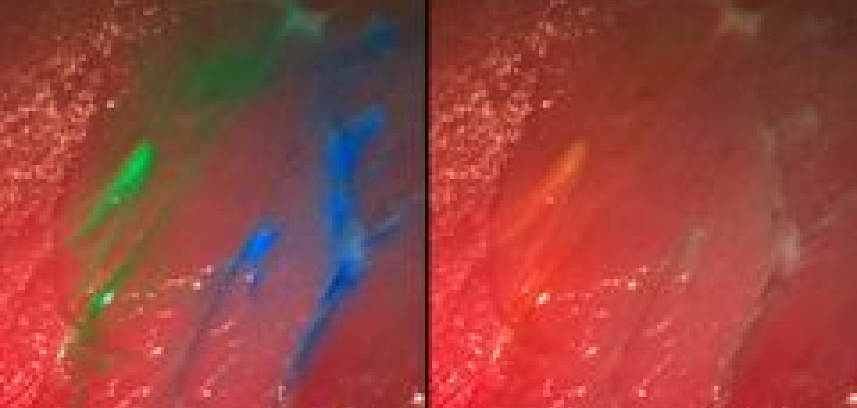
The system, developed by researchers at the Fraunhofer PAMB in Germany, which belongs to the Fraunhofer Institute for Manufacturing Engineering and Automation (IPA), can display fluorescent molecules that ‘paint’ the cancer tissue. These are injected into the patient’s blood circulation prior to the operation and selectively attach onto the tumour. If the corresponding area is then illuminated with a specific wavelength, fluorescence is emitted and the malignant tissue glows green, blue, red, or any other colour, depending on the injected dye, while the healthy tissue appears normal. In this way, the surgeon can see clusters of tumours cells that cannot be recognised by the naked eye.
The camera can display several fluorescent dyes and the reflectance image simultaneously in real time. The advantage is that arteries and delicate nerves that must not be injured during an intervention can likewise be coloured with dye. They too can then be detected with the new camera, since they are set apart from their surroundings.
Nikolas Dimitriadis, head of the Biomedical Optics Group at PAMB, said: ‘The visibility of the dye to the camera depends on the selection of the correct set of fluorescence filters. The filter separates the incident excitation wavelengths from the fluorescing wavelengths so that the diseased tissue is also set apart from its surroundings, even at very low light intensities.’
The researchers require only one camera and one set of filters for their photographs, which can present up to four dyes at the same time. Software developed in-house processes the images in seconds and presents it continuously on a monitor during surgery. The information from the fluorescent image is then superposed onto the normal colour image.
‘The operator receives significantly more accurate information. Millimetre-sized tumour remnants or metastases that a surgeon would otherwise possibly overlook are recognisable in detail on the monitor. Patients operated under fluorescent light have improved chances of survival,’ said Dimitriadis.
In order to be able to employ the multispectral fluorescence camera system as adapt ably as possible, it can be converted to other combinations of dyes. ‘One preparation that is already available to make tumours visible is 5-amino levulinic acid (5-ALA). Physicians employ this especially for glioblastomas - one of the most frequent malignant brain tumours in adults,’ explained Dimitriadis. 5-ALA leads to an accumulation of a red dye in the tumour and can likewise be detected with the camera.
The first clinical tests of this multispectral fluorescence camera system are planned for 2014, with patients suffering from glioblastomas.
In the future, it is hoped that the technology could be integrated into various medical imaging systems such as surgical microscopes and endoscopes.
