A low-cost stereovision device that could transform the way optometrists diagnose glaucoma has been developed by researchers at the University of Strathclyde.
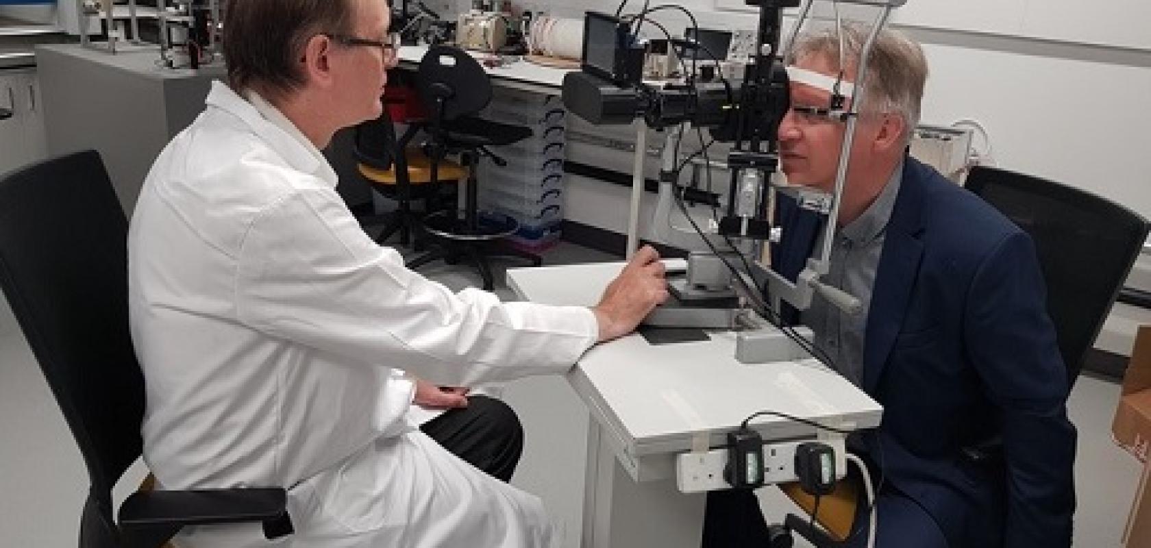
The imager is an add-on to a slit lamp, equipment commonly used by optometrists. It gives 3D images of the structure of the back of the eye, which can improve the accuracy of diagnosing eye diseases such as glaucoma.
Glaucoma is the third most common cause of visual impairment worldwide, with an estimated 7.7 million people affected. It has been shown that 3D imaging can increase diagnostic accuracy, but technology such as optical coherence tomography can be prohibitively expensive – OCT machines can cost up to £100,000.
The new technology is a simple and inexpensive add-on to a standard slit lamp, which most optometrists have access to. The device reconstructs stereovision data from images taken with low-cost cameras.
The device has been developed by Dr Mario Giardini, Dr Ian Coghill, and Kirsty Jordan at the department of biomedical engineering of the University of Strathclyde.
Dr Iain Livingstone, consultant ophthalmologist at NHS Forth Valley, who has collaborated with Dr Giardini on previous ophthalmology projects, said: 'So much of what we do as eye doctors depends on seeing things in 3D. While photographs can be helpful, this innovation uses visible light to recreate a high fidelity 3D representation of eye structures, allowing precise measurements to be taken in a completely new way, piggybacking on the method of examination we already do routinely.'
He continued: 'It’s a crucial addition to the way we interpret information, harnessing digital to glean so much more from a slit lamp exam, with potential reach far beyond the hospital toward community optometry, bringing nuanced measuring tools closer to home for patients.'
The researchers also hope it can eventually be used to detect eye cancer, with the potential to supplant ocular ultrasound for measuring solid tumours of the eye.
The technology can also be used to image the front of the eye, which is important for cornea transplant patients as many machines can’t measure the edge of the cornea.
Dr Giardini said: 'The technology has the potential to revolutionise the screening and follow-up within the community of conditions such as glaucoma, as any optometrist, anywhere in the world, could afford it. This work makes eye diagnostics more accessible, reducing inequalities.'
The initial prototyping was funded by the Engineering and Physics Research Council, part of UK Research and Innovation. The next step is to make the technology available to the medical community, and the university has partnered with IDCP, a digital innovation group, to turn it into a medical product.