Researchers have created a prototype optical scanning system based on line scan imaging instead of a traditional 2D sensor.
The device is intended to be used for monitoring hand diseases and disorders, such as eczema, as well as various skin growths, such as nevus, to monitor their size and area.
The motivation behind the development is that there is a continuous, critical need to collect the images of the human body in a precise, repeatable, and high-quality manner – whether the goal is to perform medical imaging for dermatology or wound care purposes, or to collect data for biometric applications.
The experimental scanning system, developed at the Brno University of Technology in the Czech Republic, offers flexible capability in that its field of view can be changed based on the diagnostic needs, thus enabling scanning of body parts of various sizes without having to change the physical configuration – whether the part is a finger, hand or entire arm.
Another advantage is that the system doesn't require the patient to touch the scanning surface, preventing the need to clean and disinfect it at regular intervals – a hygienic requirement for medical devices used by multiple people.
The line scan camera – a Basler raL6144-16gm with 6,144 px resolution – acquires 5,120 lines at a rate of 500 lines per second. The system uses a GigE interface and an API script was developed to handle its initialisation, data grabbing, and image construction. Triggering and frame grabbing are performed using software scripts.
The system also leverages Chromosens’ Corona II LED Illumination module as its light source, with a brightness of up to 3,500,000 lux and exceptional homogeneity. The Brno University research team selected a 190mm top light dark field module for their device with a minimum white illuminance of 800,000 lux. The module’s reflector design shapes the light in a way that eliminates chromatic aberrations.
In addition to the illumination module and line scan camera, the optical scanning device employs a linear movement system, a Chromasens XLC4-1 LED control unit, a Raspberry Pi 3 single-board computer, and a microcontroller unit. It is capable of producing 350 dpi images of the variable field of view without having to make concessions to the quality due to its mechanical properties. It allows for objects up to 120 × 43cm in size to be scanned while maintaining the 350 dpi spatial resolution.
The XLC4-1 unit is controlled over a Telnet interface. A series of simple commands provided by the unit's documentation sets the intensity of the illumination and turns the light module on and off. Commands to monitor the status of the illumination unit and the module are also utilised.
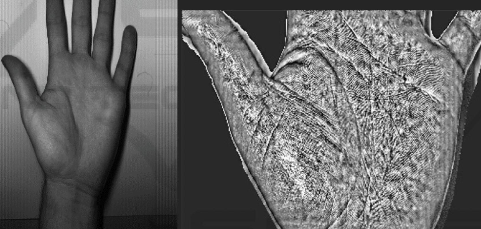
Dorsal and palmar images of hands were taken and examined for the usage in hand geometry recognition as well as performing measurements of various skin features usable for biomedical applications. The detail of the images were deemed suitable for monitoring skin diseases and disorders, as well as analysing the rate of change in the wound healing process. The device could also be used to perform recognition based on hand geometry if other biometrics are unavailable, for example, due to injuries such as a burn or skin diseases.