Scientists at the Dresden University of Technology have developed a high-speed imaging technique able to analyse cell samples 10,000 times faster than conventional methods.
The Real-Time Deformability Cytometry (RT-DC), which the team has called AcCellerator, is able to measure up to 1,000 cells per second. The scientists have founded a company, Zellmechanik Dresden, to commercialise the flow cytometry technique.
The mechanical properties of a cell give information about its health – how a cell deforms can act as a label-free biomarker to gain understanding about drug treatment effects, immune cell activation, stem cell differentiation, cancer prognosis, or the assessment of state and quality of cultured cells.
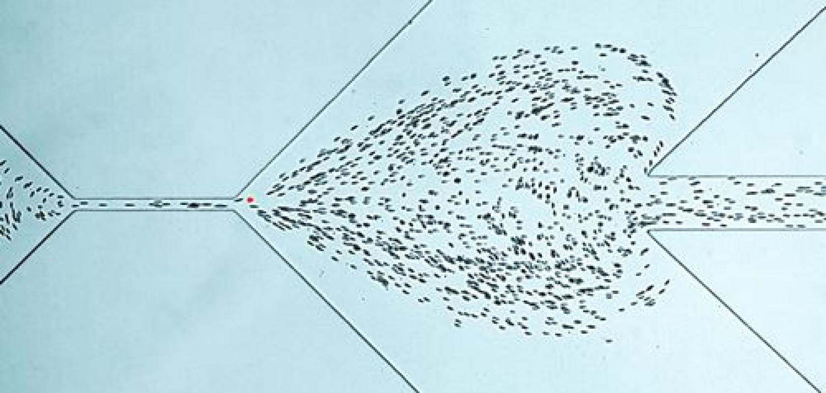
The RT-DC technique forces cells through a micro-channel, and the pressure gradient of the fluid creates a flow profile and deforms the cells. Softer cells display greater deformations.
The cells flow through the cytometry setup at a speed of 10cm/s and are viewed under a microscope with 400x magnification. An EoSens CL high-speed camera from Mikrotron is connected to the microscope and captures each individual cell, at up to 4,000 frames per second.
The camera also controls the 1μs LED light impulse sent out for each image acquisition. The standard resolution of 250 x 80 pixels is automatically adjusted to the channel width.
All images are transferred in real time to the computer via a Camera Link interface. A program based on National Instruments LabView then measures the deformation of each cell; analysing a single image takes less than 250μs.
‘This process enables us to measure the mechanical properties of several hundred cells per second. In one minute, this permits us to carry out analysis that would take a week in the technologies we used before,’ said Dr Oliver Otto, CEO of Zellmechanik Dresden. ‘Within 15 minutes, a precise characterisation of all blood cell types, including cell activation status, is analysed. Due to the high throughput of cells, only one single drop of blood is needed for the analysis.’
Thanks to the AcCellerator, cell mechanic evaluation has become usable in clinical applications for the first time. In the future, mechanical fingerprinting of cells could be used for fast diagnosis as well as for monitoring infections. Blood count changes or metastasising cells can be detected in minutes.
The technology is also opening up many new areas of application in research by enabling scientists to examine all processes in which cytoskeleton changes are responsible for the mechanical stabilisation of the cell, including migration or cell division.