A time-lapse 3D video of a zebrafish heart growing over a day has been captured for the first time by a new microscope imaging technique.
Researchers from the universities of Glasgow and Edinburgh were able to record individual heart cells growing and dividing in the beating heart over 24 hours. It is hoped the work, published in Nature Communications, will give insight into how human hearts form, grow and heal.
The fluorescence time-lapse imaging technique uses a thin sheet of laser light to build up images into a full 3D image of the heart layer by layer. The team combined this with real-time image analysis using visible light to control when to fire the lasers for imaging the heart.
While 3D images of the beating heart had been recorded in the past, a time-lapse video of the heart growing has not been possible until now.
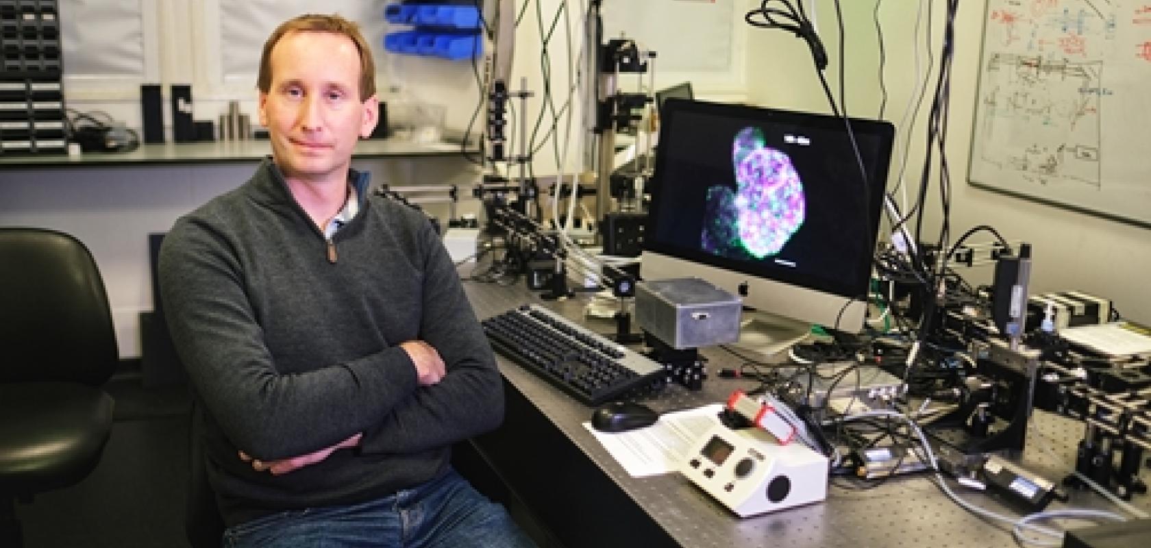
Dr Jonathan Taylor, of the University of Glasgow’s School of Physics and Astronomy, explained: ‘Zebrafish embryos’ hearts beat three times a second, and this constant motion makes them incredibly challenging targets to image accurately in three dimensions. By the time you’ve taken an image of one layer of the heart, it has moved on to another part of its cardiac cycle. To add to the challenge, heart cells can be damaged by over-exposure to laser light that is needed for fluorescence imaging.’
The team’s microscope uses visible light to image the beating heart of the zebrafish through its transparent skin. By analysing these images, laser light can then be fired selectively at the heart to capture specific moments in its cycle.
In this way, exposure to damaging laser light is minimised allowing the scientists to image the heart over the course of a day without harming the fish.
Zebrafish hearts are similar to those of humans, making them useful in cardiac research. The British Heart Foundation – one of the funders of the study – hope that the new method of visualising zebrafish hearts will provide scientists with valuable new insights into the cellular and subcellular processes which occur during the earliest stages of heart development.
Dr Martin Denvir of the University of Edinburgh, one of the paper’s co-authors, said the knowledge gained from the new technique could be used to develop treatments for abnormal heart formation in the future.
The scientists have also been able to observe immune cells travelling to injured areas of the heart, which could help guide the modification of immune response to treat cardiac inflammation and heart disease.
The paper, titled ‘Adaptive prospective optical gating enables day-long 3D time-lapse imaging of the beating embryonic zebrafish heart’, is published in Nature Communications. The research was supported with funding from the British Heart Foundation (New Horizons award), the Engineering and Physical Sciences Research Council, the Royal Society of Edinburgh.