Giorgia Marucci from Pro-Lite Technology visits the National Gallery in London to find out how hyperspectral imaging was used to reveal the history behind Leonardo da Vinci’s The Virgin of the Rocks
An image is the representation of an object or person. The ability of Leonardo da Vinci to represent reality was extraordinary. It resulted from his understanding of science, the study of light, and curiosity about the world. On the 500th anniversary of his death, the National Gallery in London is celebrating the artist with its exhibition Leonardo: Experience a Masterpiece (until 26 January 2020), where visitors can discover and rediscover one of the most famous of da Vinci’s pieces hung in the gallery, the Virgin of the Rocks. The show brings together findings from research, including an investigation using imaging techniques performed by the scientific and conservation departments of the National Gallery.
A problematic commission
In 1483 the Confraternity of the Immaculate Conception of the Virgin of San Francesco Grande church in Milan commissioned da Vinci, and the de Predis brothers, to paint the central panel of the altarpiece of the Chapel of the Immaculate Conception. Da Vinci completed a first version of the painting in about 1485 but, thinking that the fee offered by the Confraternity was lower than the painting’s true worth, ended up selling it to another buyer. Today, this earlier version of the Virgin of the Rocks hangs in the Louvre in Paris.
In the early 1490s, da Vinci started work on another version of the same subject, thought to have been delivered by 1499. However, with his customer still unhappy with the finish, da Vinci kept working on this second painting – the one now owned by the National Gallery – between 1506 and 1508. It was only then that he received final payment, 25 years after starting the first version. At this point, da Vinci had a different perception of the world, a fuller understanding of the behaviour of light and his artistic ability, which makes the two works of art distinct. (For further details on the historical context, see the exhibition catalogue, L. Kharibian, Leonardo: Experience a Masterpiece, London, 2019.)
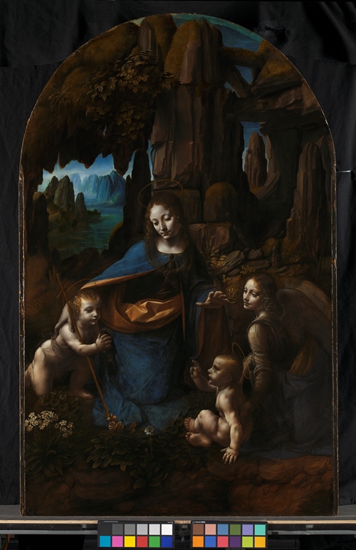
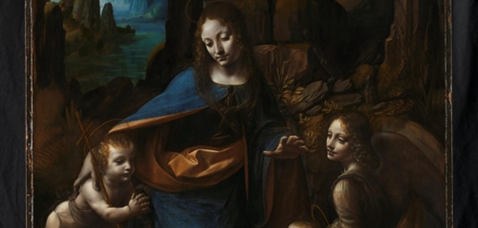
Leonardo da Vinci, 1452–1519, The Virgin with the Infant Saint John the Baptist adoring the Christ Child accompanied by an Angel (The Virgin of the Rocks) about 1491/2-9 and 1506-8. Oil on poplar, thinned and cradled. 189.5 x 120cm. Copyright: The National Gallery, London
Unexpected discoveries
To better understand da Vinci’s modus operandi and his creative process, the National Gallery’s scientific and conservation departments made a number of studies on the Virgin of the Rocks, using non-invasive and non-destructive imaging techniques.
X-ray radiography – the same as that used in medicine – was first performed in 1948, and then later in 2005 with more advanced technology. In both cases, it showed little information concerning the painting itself, the image being dominated by the cradled panel.
New research was conducted in 2005 and 2006 using infrared reflectography, an imaging method that relies on the reflective and absorptive proprieties of materials when illuminated by an infrared light source. Paint layers are usually transparent in this wavelength region, while carbon-based materials, such as the charcoal used to make a preparatory sketch hidden underneath the paint, absorb in the infrared. Traces of the underdrawings for the Virgin of the Rocks came to light in this study thanks to the infrared reflectogram. Surprisingly, however, these images also showed lines referring to a different composition. This unexpected drawing shows a different figure of the Virgin Mary, with her head in a 3/4 pose, possibly turned toward the child who is unfortunately not visible in the infrared image, and her left hand brought to the chest, her right arm outstretched to the side.
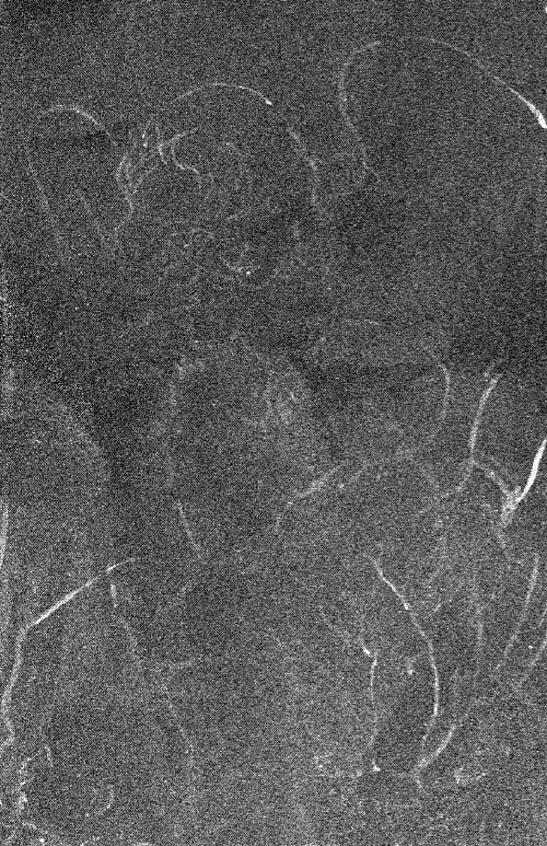
Figure 1: Macro XRF map showing distribution of zinc revealing angel and baby of the first composition (under the landscape at the right side of the painting). Copyright: The National Gallery, London
Fortunately, technology evolves, and this year a new study was performed to find out more about these underdrawings and a design that looks so different from the final composition known today. The analysis included macro x-ray fluorescence (XRF) scanning and hyperspectral imaging. The macro XRF output are maps of the distribution of chemical elements at and below the surface of the painting. Unexpectedly, the distribution map of the element zinc highlighted the figures of an angel and child on the right-hand side of the painting (figure one), which the infrared reflectogram in 2005 had not shown.
Hyperspectral imaging reveals more
Hyperspectral imaging was used to try to obtain a clearer and more complete image of the underlying abandoned composition. The hyperspectral acquisitions were performed with an imaging system built in-house that operates in the shortwave infrared range (1,000nm to 2,500nm). This is a push-broom hyperspectral camera that uses a grating spectrometer with a 30µm width slit for wavelength dispersion. An intermediate relay lens projects the dispersed spatial line onto a 1,280 x 1,024-pixel InSb detector, providing a 3nm spectral sampling interval and 500 spectral bands. The area under study was illuminated with two 125W halogen lamps controlled by a rheostat.
Under the working conditions used for studying the painting, the optics of the system provided a field of view of about 21cm. Since the painting measures 189.5 x 120cm, 11 scans were required to cover its entire surface. Each scan included a 15 per cent region of overlap to be able to mosaic the whole surface. The scans were acquired by moving the painting – installed on a computer-controlled micro-positioning easel – and keeping the camera static. With an integration time of 100ms for image acquisition, each of the 11 hyperspectral datacubes took 15 minutes to generate.
Hyperspectral imaging is a powerful technique that provides the best performance after spectral data treatment. The raw data sets were flat-field corrected with a white Labsphere Spectralon diffuse reflectance target panel (99 per cent), darkfield corrected, and calibrated with a black and a white standard, which were included in the field of view at the acquisition time.
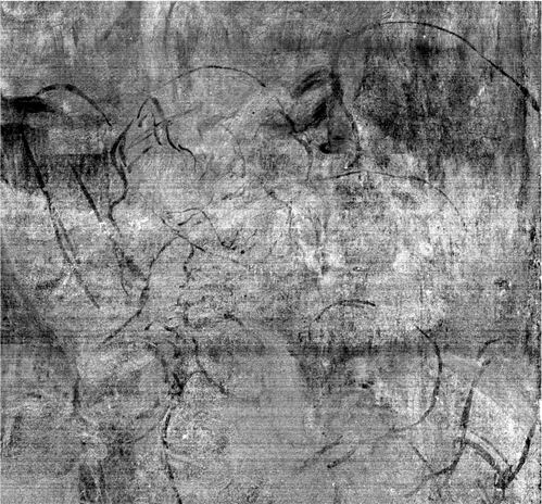
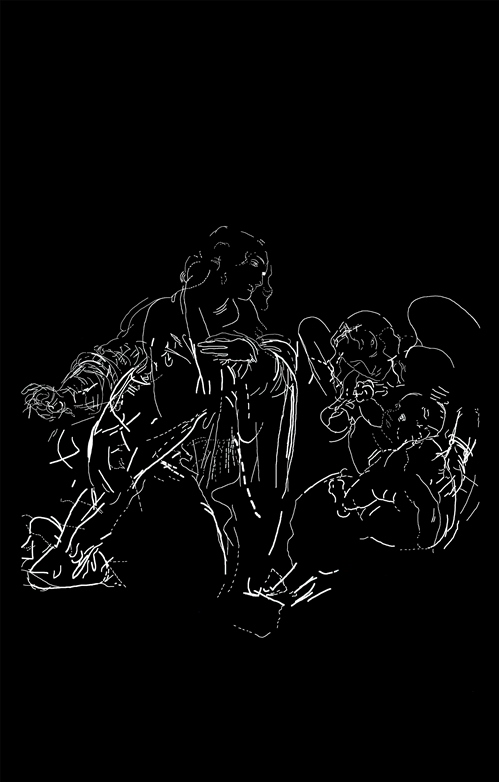
Figure 2: Detail from hyperspectral imaging data, revealing the drawing for the angel and baby of the first composition (under the landscape at the right side of the painting). Copyright: The National Gallery, London
To recreate the entire surface, the 11 scans were mosaiced together. The interpretation of the data was performed by visualising single-band images and creating false-colour images, assigning to the red, green and blue channels three spectral bands of interest; the false-colour image highlights differences in the painting materials that otherwise could not be seen.
Another approach to identify interesting features relative to the spectral behaviour of the materials is to apply chemometrics, in this case principal component (PC) and minimum noise fraction (MNF) transforms. Dr Marta Melchiorre, who performed the analysis, found that MNF was particularly effective in providing images with improved signal-to-noise ratio, which highlighted features not visible in the original images. Figure two shows one of the eigen images resulting from the MNF transform and highlights the traces of the angel and the child outlines, matching with the zinc map (figure one).
The strength of hyperspectral imaging goes beyond making the invisible visible. The possibility to extract reflectance spectra from each pixel of the image allows pigments to be identified, complementing XRF results. For example, it is possible to identify a copper-based blue as azurite.
Tracing of the lines relating to underdrawings for the first composition, incorporating information from all technical images. Copyright: The National Gallery, London
To be continued...
Imaging techniques are a suitable and effective approach to investigate pieces of art, allowing the study of a whole work in a non-invasive and non-destructive manner. The research done by Dr Marta Melchiorre and her colleagues at the National Gallery demonstrates how hyperspectral imaging can reveal hidden underdrawings made in the early creation stages of the painting. The imaging method can also identify the composition and map materials over the surface of the painting, saving time when compared to point-by-point spectroscopic techniques.
The evolution of hyperspectral imaging means that more meaningful data can be collected compared to older methods like infrared reflectography, providing a deeper understanding of the painting. The possibility to process the spectral data with multivariable statistics gives information that goes beyond the appearance of the image. As an ongoing project, further analysis of the data could still reveal more.
Finally, the interpretation of findings is ongoing and further work is planned to fully understand the results within the historical context of this painting.
Acknowledgements
The author would like to thank Dr Marta Melchiorre, Dr Catherine Higgitt and Marika Spring from the scientific department of the National Gallery for the support and the time dedicated to providing an exclusive insight into Leonardo da Vinci’s work.
About the author
Dr Giorgia Marucci is an applications specialist working in the spectroscopy and spectral imaging team at Pro-Lite Technology. Pro-lite Technology is a supplier of specialist equipment and services with a technical focus in photonics, in instruments for measuring light and the optical properties of materials, and in optical spectroscopy and spectral imaging. For further details, please visit http://pro-lite.co.uk/File/hyperspectral_imaging.php.

Further reading by the author
The Art of Raman Spectroscopy - Pro-Lite’s Dr Giorgia Marucci discusses the application of Raman spectroscopy in the cultural heritage field
Write for us
If you use hyperspectral imaging and would like to write about a system you developed and deployed successfully, please get in touch: greg.blackman@europascience.com