Cardiac doctors have trialled a 3D holographic imaging system as an aid in minimally invasive heart procedures. Jessica Rowbury speaks to Dr Elchanan Bruckheimer, a paediatric cardiologist who presented the results of the trial at a recent cardiovascular symposium
Doctors working in collaboration with the Schneider Children’s Medical Center in Petach Tikva, Israel have completed a clinical trial showing how 3D holographic images can be used to guide minimally invasive heart procedures. The results of the study were presented on 29 October at the Transcatheter Cardiovascular Therapeutics (TCT) scientific symposium in San Francisco.
The holographic imaging system was developed by Royal Philips, the healthcare arm of Philips, and holographic display company, RealView Imaging. It is hoped that the technology will be available for use in clinical catheter laboratories in the next two years, according to Dr Elchanan Bruckheimer, paediatric cardiologist and director of the Cardiac Catheterization Laboratories at Schneider Children’s Medical Center, who presented the results at the symposium.
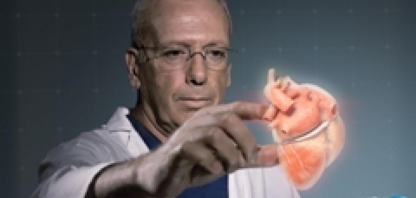
Eight patients were involved in the trial, which used RealView’s visualisation technology to display interactive, real-time 3D holographic images acquired by Philips’ X-ray and cardiac ultrasound systems. Doctors in the team were able to view detailed dynamic 3D holographic images of the heart 'floating in free space' during a minimally-invasive structural heart disease procedure.
‘The study was carried out to demonstrate the technical feasibility of producing holograms for the first time in a catheter laboratory setting, in a way that would be useful to the doctor by allowing them to interact with the hologram,' explained Dr Bruckheimer.
After successful results from the pilot study, this technology is not far from becoming available in a clinical setting, as Dr Bruckheimer explained: ‘Hopefully within the next two years we will be able to present a commercial working device that can be put into any major catheter lab and be put to use.'
During minimally invasive heart procedures, doctors use x-ray imaging, which provides visualisation of catheters and heart implants, and ultrasound imaging, providing detailed insights into the heart’s soft tissue anatomy. Typically, doctors will look at a 60-inch high definition screen during the procedure to see 3D images on a 2D screen.
Steve Klink, director of communications and senior press officer at Philips Group Communications, said: ‘The idea was to combine our ultrasound and x-ray scanners and, instead of just presenting the data on a standard 2D screen, to also present that data as a hologram. The 3D hologram technology is capable of translating the 3D datasets from our scanners into a real life hologram of the patient’s heart.’
Doctors were able to manipulate the projected 3D heart structures by touching the holographic volumes in front of them.
‘The holographic projections enabled me to understand and interrogate the 3D spatial anatomy of the patient’s heart, as well as to navigate and appreciate the device-tissue interaction during the procedure,’ said Dr Einat Birk, paediatric cardiologist and Director of the Institute of Pediatric Cardiology at Schneider Children’s Medical Center.
Dr Bruckheimer added: ‘The ability to reach into the image and apply markings on the soft tissue anatomy in the x-ray and 3D ultrasound images would be extremely useful for guidance of these complex procedures.’
Klink explained: ‘Potentially, with this new technology, the doctor can have all the benefits of open heart surgery, in that he can touch the heart, see the features of the heart, and manipulate it, but it will be a minimally invasive procedure that will have less impact on the patient.’
Image-guided therapies for heart diseases have greatly increased the need for live 3D image guidance, to supplement today’s live 2D image guidance. ‘Medicine is moving so that procedures such as micro valve repair, valve replacements and complex electrophysiological procedures such as catheter ablation therapy for heart arrhythmias are carried out in the catheter lab,’ said Dr Bruckheimer.
According to Klink, an important trend in healthcare is the transition from surgical procedures to more minimally-invasive procedures. ‘We think that technologies like this could potentially drive the trend from surgery to minimally invasive procedures to allow for faster patient recovery, shorter hospital stays, and fewer complications,’ he said.