A European project led by the UK's University of Sheffield will develop image-based biophysical models of the human brain to enable early diagnosis of dementia and help doctors predict the progression of the disease.
The university says the model will be the first to combine a wide range of physiological data from medical imaging – such as blood flow, brain tissue properties and cellular activity – with psychological measures such as memory and cognitive function. It will also bring in demographic, genetic, lifestyle and environmental factors, making it much more sensitive than existing diagnostic tools.
The aim is to develop a way for doctors to identify at an early stage – before obvious symptoms appear – not only whether a patient has or is developing dementia, but which form of dementia it is. Diagnosis of the most common form of dementia – Alzheimer’s – in the UK still takes an average of 32 months after symptoms are first noticed, compared to 20 months in Europe as a whole.
'We currently rely on basic cognitive tests and conventional brain scans for diagnosis of dementia but these aren’t good enough to recognise the disease at an early stage,' explained the project’s scientific director, professor Alex Frangi, from the university’s faculty of engineering.
'If we can identify a much wider range of markers which provide better and earlier diagnosis, then that gives doctors time to delay the progression of the disease. This would be not only improving patients’ quality of life, but it would also reduce the burden on carers and the enormous costs of supporting people with dementia.'
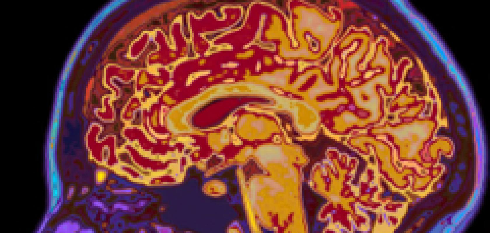
The engineers, scientists and clinicians will draw on historical data from over 20,000 patients across Europe to build their model. In addition, three studies will gather new data from 150 patients to test hypotheses on contributory factors for the disease. One of these studies will take place in Sheffield and will involve a new brain imaging technique that can identify changes in the elasticity and rigidity of brain tissue.
Iain Wilkinson, Professor of Magnetic Resonance (MR) Physics in the university’s faculty of medicine, dentistry and health, said: 'The temporal lobes, part of the brain affected in Alzheimer’s, lose tissue as the disease progresses. Their rigidity or ‘softness’ is likely to alter as part of this process before the tissue loss occurs.
'A technique called MR elastography, developed to scan for these properties in liver tissue, is being adapted by colleagues in the UK and Switzerland for use in the brain. We’ll be applying this technique with patients in Sheffield, to see if it could be used in the model to help earlier diagnosis.'
The project is one of several underway at the University of Sheffield aiming to create a ‘virtual physiological human’ – a computer model of a patient which can be personalised to test potential treatments. The University’s INSIGNEO Institute for In-Silico Medicine is one of the major European hubs for this field of research.
