Work to develop an imaging technique to measure blood flow across the surface of the brain that could help patients undergoing neurosurgery, has received a four-year, $1.8 million grant from the National Institutes of Health.
Biomedical engineering professor Andrew Dunn and his team at the Cockrell School of Engineering at The University of Texas at Austin are developing a laser-based technique, called multi-exposure speckle imaging (MESI), for measuring blood flow during brain surgery or after a stroke.
The goal of the NIH grant is to enable UT Austin researchers to get real-time blood flow measurements, allowing neurosurgeons to better assess problem areas by distinguishing healthy arteries from blocked arteries. For the past five years, MESI has been studied in humans during surgery and has progressed to deliver clinically useful images, but it is still difficult to give precise blood flow measurements of the brain to surgeons.
‘We hope to transform the images we have now and to be able to give surgeons quantitative data on blood flow that we can provide in real time, with no dyes and no interruptions to the surgery,’ Dunn said.
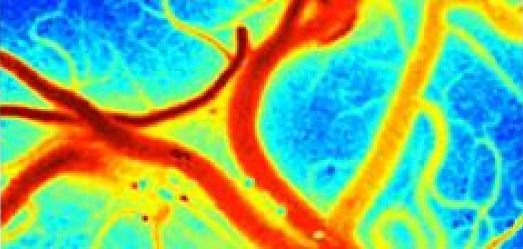
The MESI technique uses a laser expanded to illuminate the entire surgical area at once. When the laser reflects off of tissue, it provides a grainy image or speckle pattern. The technique analyses the fluctuations in the speckles and converts that information into a motion map, similar to a weather map, which identifies blood flow and blocked arteries. Another major advantage is that the MESI device can be added to existing surgical microscopes, so it does not require entirely new equipment.
Today, when patients undergo surgery for an aneurysm – a bulge on the wall of an artery – they are first injected with a fluorescent dye that lights up perfused vessels and darkens blocked vessels in the brain. Neurosurgeons use this imaging as a guide to determine where to clip an artery. But vessels often have varying degrees of blood flow, and current imaging methods do not quantitatively measure blood flow. Current methods are also limited if patients are allergic to dyes and because a surgeon is able to use the dye method only once or twice during a surgery.
In addition to neurosurgery, the researchers believe that MESI has the potential to provide quantitative imaging – real-time measurements of blood flow that allow researchers to see any damage caused by a stroke and how the brain may be healing.
In the days and weeks after a stroke, the brain undergoes changes, growing new vessels to repair itself. Little is known about this remodelling process, but MESI may be able to provide measurements that could be helpful to drug developers in the design of drugs to restore blood flow to the brain.
By the end of the four-year grant, Dunn and his team hope to have both software and a tool ready for commercialisation.
Dunn and his lab are collaborating with Dr Douglas Fox, a neurosurgeon and executive medical director at NeuroTexas Institute at St. David's HealthCare, to translate the technology to the clinical setting.
Further information:
Cockrell School of Engineering at The University of Texas at Austin
