Queen Mary University and ZEISS test super resolution imaging technique for DNA repair
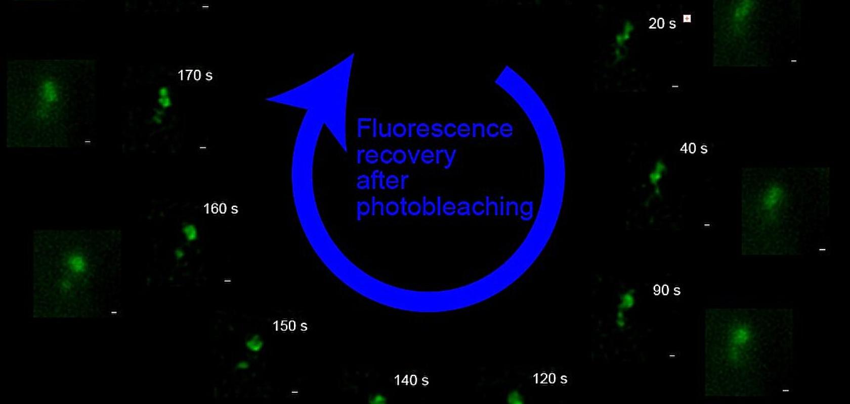
FRAP-SR tracks foci through time in seconds. The fast reappearance of the green signal after photobleaching indicates fast exchange of the protein (Image: Queen Mary University of London)
A new microscopy technique developed by Queen Mary University has achieved live-cell imaging at 60nm, and could be used to repair DNA
Register for FREE to keep reading
Join 10,000+ vision professionals driving innovation in automation, AI and imaging with:
- Expert insights on vision, robotics, AI & embedded tech
- Newsletters and features covering the full imaging landscape
- Visionaries series: leadership strategies in imaging
- Free panels on smart manufacturing & autonomy
- White Papers & updates for smarter integration
Sign up now
Already a member? Log in here
Your data is protected under our privacy policy.