48-camera microscope developed for high-resolution 3D surgical imagery
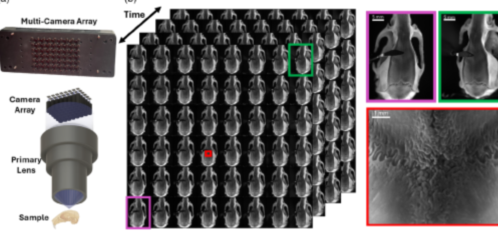
(L-R): Multicamera FiLM-Scope array with 3D model of FiLM-Scope, 48x ex-vivo rat skull snapshots, the angular range of the system (purple and green) and the high resolution (red) (Image: Duke University)
A Fourier lightfield multiview microscope has been developed by Duke University, opening up 3D depth sensing for microsurgery with 11µm precision
Register for FREE to keep reading
Join 10,000+ vision professionals driving innovation in automation, AI and imaging with:
- Expert insights on vision, robotics, AI & embedded tech
- Newsletters and features covering the full imaging landscape
- Visionaries series: leadership strategies in imaging
- Free panels on smart manufacturing & autonomy
- White Papers & updates for smarter integration
Sign up now
Already a member? Log in here
Your data is protected under our privacy policy.